A Case of Nasal NK T-Cell Lymphoma Presenting With Persistent Epiphora
- Affiliations
-
- 1Department of Otorhinolaryngology-Head and Neck Surgery, Dong-A University College of Medicine, Busan, Republic of Korea
- 2Department of Pathology, Dong-A University College of Medicine, Busan, Republic of Korea
- KMID: 2536588
- DOI: http://doi.org/10.18787/jr.2022.00415
Abstract
- Nasal type natural killer/T-cell lymphoma (NNKTL) is a rare and aggressive subtype of non-Hodgkin lymphoma originating from a natural killer cell or γδ T cell infected by the Epstein-Barr virus. It usually invades the aerodigestive tract and can rapidly destroy the paranasal sinus, hard palate, and central nervous system. NNKTL is often mistaken for benign conditions such as chronic hypertrophic rhinosinusitis or mucosal inflammatory change, as endoscopic findings of NNKTL presenting nasal mucosal hypertrophy are similar to endoscopic findings for these abovementioned benign conditions. Here, the authors report the diagnosis and examination of NNKTL in a 58-year-old male patient who visited our clinic for nasal cavity discomfort after he underwent a dacryocystorhinostomy to treat dacryocystitis.
Keyword
Figure
-
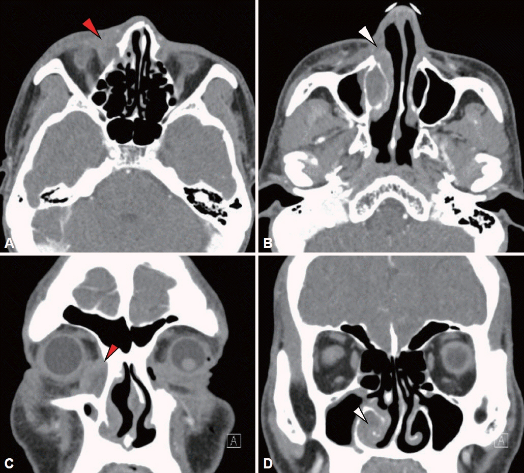
Fig. 1. Radiologic findings. Paranasal CT shows inhomogenous enhancement and soft tissue thickening around right nasolacrimal duct (red arrowheads). A: Axial view. B: Coronal view. Paranasal CT shows mucosal thickening of right inferior turbinate and inferior meatus (white arrowheads). C: Axial view. D: Coronal view.
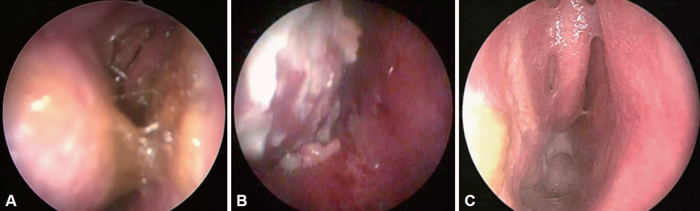
Fig. 2. Endoscopic findings. A: Shows profuse mucus discharge and swelling of right inferior turbinate adhere to right septal wall mucosa. B: Shows mucopus discharge and adhesion of right inferior turbinate to its adjacent structures. Whitish plaques are newly found on the surface of inferior turbinate. C: Shows no evidence of recurrence. There are no inflammatory change or hypertrophy of nasal mucosa.
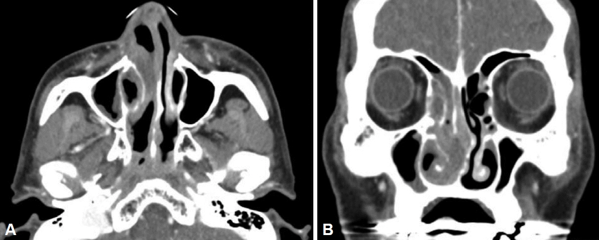
Fig. 3. Radiologic findings. Paranasal CT (1 month after Fig. 1) shows nonenhanced mucosal thickening of osteomeatal unit, right maxillary sinus, base of nasal cavity and nasal septum. Thickening of mucosa remarkably progressed compared to CT 1 month ago (A: Axial view, B: Coronal view).
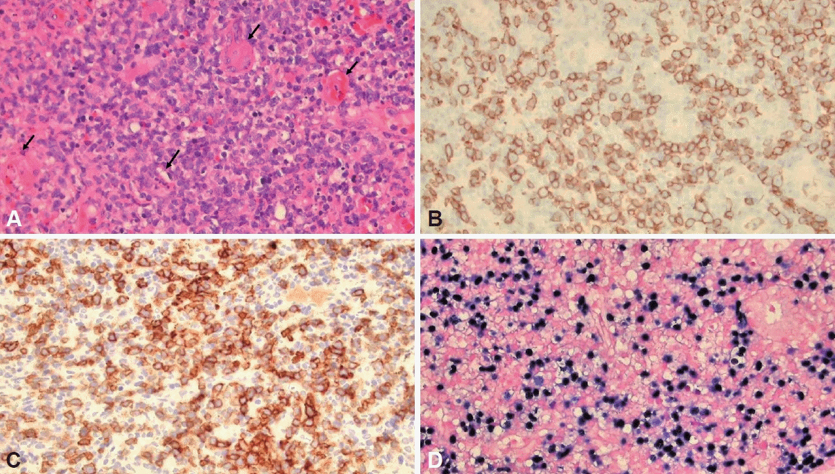
Fig. 4. Pathologic findings. A: The tumor was composed of medium-sized lymphoid cells with irregularly folded nuclei and showed angiodestructive growth pattern (arrows) (hematoxylin eosin stain, ×400 magnification). B: Tumor cells showed membranous positivity for CD56 immunohistochemical stain (CD56, ×400 magnification). C: Tumor cells were also positive for CD30 immunohistochemical stain (CD30, ×400 magnification). D: In situ hybridization for Epstein-Barr virus (EBV)-encoded small RNA (EBER) demonstrated the presence of EBV (EBER, ×400 magnification).
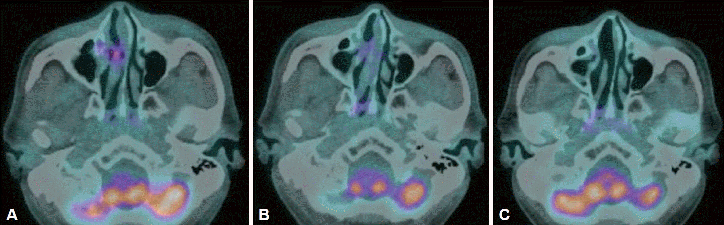
Fig. 5. Radiologic findings of positive emission tomograpy-CT. A: Hypermetabolic lymphoma in right middle meatus, middle turbinate and inferior turbinate. B: 2 month after (A) shows diffuse mild hypermetabolic mucosal thickening of right nasal cavity. Focal hypermetabolic thickening of right nasal cavity was disappeared. C: 3 month after (B). Diffuse mild hypermetabolic lesion was disappeared and there are no abnormal hypermetabolic uptake.
Reference
-
References
1. Hung LY, Chang PH, Lee TJ, Hsu YP, Chen YW, Fu CH, et al. Extranodal natural killer/T-cell lymphoma, nasal type: clinical and computed tomography findings in the head and neck region. Laryngoscope. 2012; 122(12):2632–9.
Article2. Harabuchi Y, Takahara M, Kishibe K, Nagato T, Kumai T. Extranodal natural killer/T-cell lymphoma, nasal type: basic science and clinical progress. Front Pediatr. 2019; 7:141.
Article3. Tse E, Kwong YL. The diagnosis and management of NK/T-cell lymphomas. J Hematol Oncol. 2017; 10(1):85.
Article4. Wang H, Xia X, Qian C. Extranodal natural killer/T cell lymphoma, nasal type in the middle cranial fossa: a case report. Medicine (Baltimore). 2018; 97(34):e12028.5. Ko YH, Kim CW, Park CS, Jang HK, Lee SS, Kim SH, et al. REAL classification of malignant lymphomas in the Republic of Korea: incidence of recently recognized entities and changes in clinicopathologic features. Hematolymphoreticular Study Group of the Korean Society of Pathologists. Revised European-American lymphoma. Cancer. 1998; 83(4):806–12.6. Lee SS, Cho KJ, Kim CW, Kang YK. Clinicopathological analysis of 501 non-Hodgkin’s lymphomas in Korea according to the revised European-American classification of lymphoid neoplasms. Histopathology. 1999; 35(4):345–54.
Article7. Jiang M, Zhang L, Xie L, Zhang H, Jiang Y, Liu WP, et al. A phase II prospective study of the “Sandwich” protocol, L-asparaginase, cisplatin, dexamethasone and etoposide chemotherapy combined with concurrent radiation and cisplatin, in newly diagnosed, I/II stage, nasal type, extranodal natural killer/T-cell lymphoma. Oncotarget. 2017; 8(30):50155–63.
Article8. Haverkos BM, Pan Z, Gru AA, Freud AG, Rabinovitch R, Xu-Welliver M, et al. Extranodal NK/T cell lymphoma, nasal type (ENKTLNT): an update on epidemiology, clinical presentation, and natural history in North American and European cases. Curr Hematol Malig Rep. 2016; 11(6):514–27.
Article9. Gill H, Liang RH, Tse E. Extranodal natural-killer/t-cell lymphoma, nasal type. Adv Hematol. 2010; 2010:627401.
Article10. Kwong YL. The diagnosis and management of extranodal NK/T-cell lymphoma, nasal-type and aggressive NK-cell leukemia. J Clin Exp Hematop. 2011; 51(1):21–8.
Article11. McBride P. Photographs of a case of rapid destruction of the nose and face. 1897. J Laryngol Otol. 1991; 105(12):1120.12. Shams PN, Selva D. Acute post-operative rhinosinusitis following endonasal dacryocystorhinostomy. Eye (Lond). 2013; 27(10):1130–6.
Article
- Full Text Links
-
- Actions
-
Cited
- CITED
-
- Close
- Share
-
- Similar articles
-
- A Case of Extranodall NK/T-cell Lymphoma, Nasal type
- A Case of Primary Nasal CD56+ NK/T cell Lymphoma with Cutaneous Involvement
- A Case of Nasal CD56+ NK/T Cell Lymphoma Mimicking Cellulitis which Developed after Persistent Orbital Swelling
- Extranodal NK/T Cell Lymphoma, Nasal Type that Occurred in Patients with Atrophic Rhinitis
- A Case of Nasal Type NK/T-cell Lymphoma
