J Korean Neurosurg Soc.
2021 Jan;64(1):136-141. 10.3340/jkns.2019.0253.
Novel Noncrossing Y-Stent Technique Using Tapered Proximal End of a Solitaire AB Stent for Coil Embolization of Wide-Neck Bifurcation Aneurysms
- Affiliations
-
- 1Department of Neurosurgery, Daejeon-Chungnam Regional Cerebrovascular Center, Chungnam National University Hospital, Chungnam National University Medical School, Daejeon, Korea
- KMID: 2510768
- DOI: http://doi.org/10.3340/jkns.2019.0253
Abstract
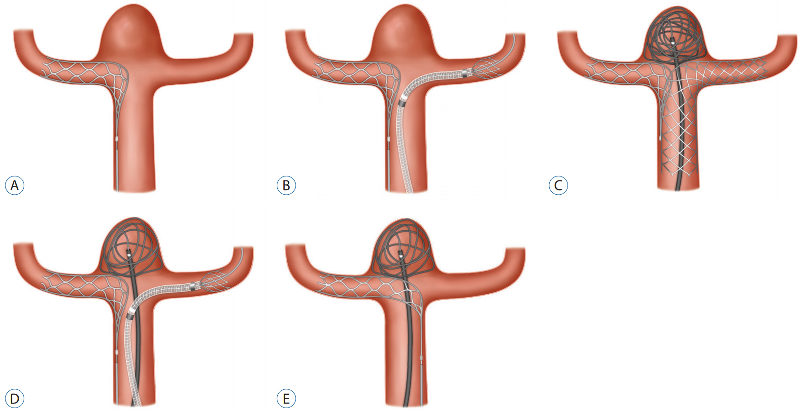
- The crossing Y-stent method is one of the indispensable techniques to achieve sufficient neck coverage during coil embolization of bifurcation aneurysms with a wide neck and/or branch incorporation. However, the inevitable hourglass-like expansion of the second stent at the crossing point can result in insufficient vessel wall apposition, reduced aneurysm neck coverage, delayed endothelialization, and subsequent higher risks of acute or delayed thrombosis. It also interferes with engagement of the microcatheter into the aneurysm after stent installation. We expected to be able to reduce these disadvantages by installing a noncrossing type Y-stent using the Solitaire AB stent, which is fully retrievable with a tapered proximal end. Here we report the techniques and two successful cases.
Keyword
Figure
Cited by 1 articles
-
Long-Term Outcomes of Placement of a Single Transverse Stent through the Anterior Communicating Artery via the Nondominant A1 in Coil Embolization of Wide-Necked Anterior Communicating Artery Aneurysms
Seung Pil Ban, O-Ki Kwon, Young Deok Kim
J Korean Neurosurg Soc. 2022;65(1):40-48. doi: 10.3340/jkns.2021.0191.
Reference
-
References
1. Brassel F, Melber K, Schlunz-Hendann M, Meila D. Kissing-Y stenting for endovascular treatment of complex wide necked bifurcation aneurysms using Acandis Acclino stents: results and literature review. J Neurointerv Surg. 8:386–395. 2016.
Article2. Cagnazzo F, Limbucci N, Nappini S, Renieri L, Rosi A, Laiso A, et al. Y-stent-assisted coiling of wide-neck bifurcation intracranial aneurysms: a meta-analysis. AJNR Am J Neuroradiol. 40:122–128. 2019.
Article3. Cho YD, Park SW, Lee JY, Seo JH, Kang HS, Kim JE, et al. Nonoverlapping Y-configuration stenting technique with dual closed-cell stents in wide-neck basilar tip aneurysms. Neurosurgery. 70(2 Suppl Operative):244–249. 2012.
Article4. Granja MF, Cortez GM, Aguilar-Salinas P, Agnoletto GJ, Imbarrato G, Jaume A, et al. Stent-assisted coiling of cerebral aneurysms using the Y-stenting technique: a systematic review and meta-analysis. J Neurointerv Surg. 11:683–689. 2019.
Article5. Klisch J, Clajus C, Sychra V, Eger C, Strasilla C, Rosahl S, et al. Coil embolization of anterior circulation aneurysms supported by the Solitaire AB Neurovascular Remodeling Device. Neuroradiology. 52:349–359. 2010.
Article6. Kono K, Shintani A, Yoshimura R, Okada H, Tanaka Y, Fujimoto T, et al. Triple antiplatelet therapy with addition of cilostazol to aspirin and clopidogrel for Y-stent-assisted coil embolization of cerebral aneurysms. Acta Neurochir (Wien). 155:1549–1557. 2013.
Article7. Kono K, Terada T. Feasibility of insertion of a microcatheter through a Y-stent in coil embolization of cerebral aneurysms and its detailed geometry by micro-computed tomography. Acta Neurochir (Wien). 156:39–43. 2014.
Article8. Samaniego EA, Mendez AA, Nguyen TN, Kalousek V, Guerrero WR, Dandapat S, et al. LVIS Jr device for Y-stent-assisted coil embolization of wide-neck intracranial aneurysms: a multicenter experience. Interv Neurol. 7:271–283. 2018.
Article9. Spiotta AM, Derdeyn CP, Tateshima S, Mocco J, Crowley RW, Liu KC, et al. Results of the ANSWER trial using the PulseRider for the treatment of broad-necked, bifurcation aneurysms. Neurosurgery. 81:56–65. 2017.
Article10. Thorell WE, Chow MM, Woo HH, Masaryk TJ, Rasmussen PA. Y-configured dual intracranial stent-assisted coil embolization for the treatment of wide-necked basilar tip aneurysms. Neurosurgery. 56:1035–1040. discussion 1035-1040. 2005.11. Ulfert C, Pfaff J, Schönenberger S, Bösel J, Herweh C, Pham M, et al. The pCONus device in treatment of wide-necked aneurysms : technical and midterm clinical and angiographic results. Clin Neuroradiol. 28:47–54. 2018.
Article12. Xue G, Zuo Q, Duan G, Zhang X, Zhao R, Li Q, et al. Dual stent-assisted coil embolization for intracranial wide-necked bifurcation aneurysms: a single-center experience and a systematic review and meta-analysis. World Neurosurg. 126:e295–e313. 2019.
Article
- Full Text Links
- Actions
-
Cited
- CITED
-
- Close
- Share
- Similar articles
-
- Waffle-Cone Technique Using Solitaire AB Stent
- Clinical and Angiographic Outcomes of Wide-necked Aneurysms Treated with the Solitaire AB Stent
- Solitaire AB Stent-Assisted Coiling of Wide-Neck Micro Aneurysms
- Outcomes of Stent-assisted Coil Embolization of Wide-necked Intracranial Aneurysms Using the Solitaire(TM) AB Neurovascular Remodeling Device
- Preliminary Results of Y-Stent-Assisted Coil Embolization of Wide-Necked Intracranial Aneurysms: 8 Consecutive Patients


