Clin Endosc.
2018 Sep;51(5):500-501. 10.5946/ce.2018.061.
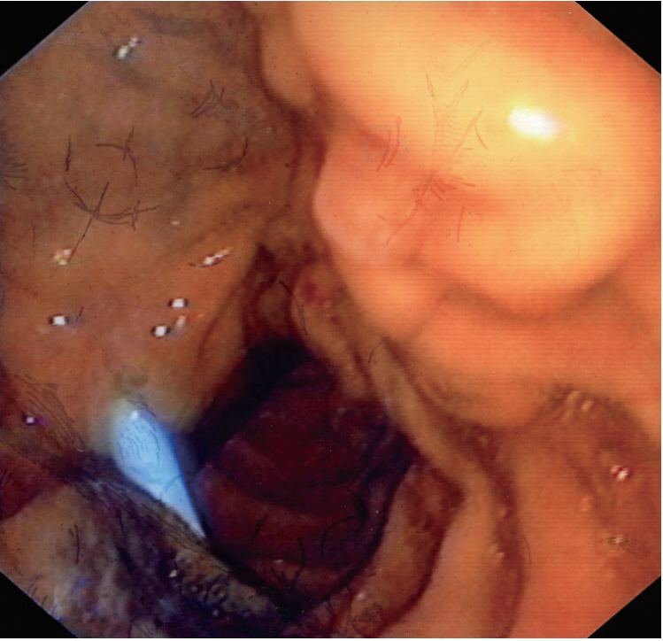
Ocular Melanoma Recurrence Presenting as Cholestatic Jaundice due to Periampullary Area Metastases
- Affiliations
-
- 1Section of Gastroenterology, Second Department of Internal Medicine Ippokration Hospital, Thessaloniki, Greece. anassot@yahoo.gr
- KMID: 2427726
- DOI: http://doi.org/10.5946/ce.2018.061
Abstract
- No abstract available.
Figure
Reference
-
1. Schuchter LM, Green R, Fraker D. Primary and metastatic diseases in malignant melanoma of the gastrointestinal tract. Curr Opin Oncol. 2000; 12:181–185.
Article2. Blecker D, Abraham S, Furth EE, Kochman ML. Melanoma in the gastrointestinal tract. Am J Gastroenterol. 1999; 94:3427–3433.
Article3. Marks JA, Rao AS, Loren D, Witkiewicz A, Mastrangelo MJ, Berger AC. Malignant melanoma presenting as obstructive jaundice secondary to metastasis to the ampulla of Vater. JOP. 2010; 11:173–175.4. Kadakia SC, Parker A, Canales L. Metastatic tumors to the upper gastrointestinal tract: endoscopic experience. Am J Gastroenterol. 1992; 87:1418–1423.
- Full Text Links
- Actions
-
Cited
- CITED
-
- Close
- Share
- Similar articles
-
- A case of Methimazole-Induced Cholestatic Jaundice With Agranulocytosis
- Four cases of ticlopidine-induced cholestatic hepatitis
- A Case of Methimazole-Induced Cholestatic Jaundice with Steroid Therapy
- A Case of Concurrent Cholestatic Jaundice and Hemolytic Anemia Due to Epstein-Barr Virus Infection
- A Case of Steven-Johnson Syndroe Associated with Cholestatic Hepatitis


