Optimal scan delay depending on contrast material injection duration in abdominal multi-phase computed tomography of pancreas and liver in normal Beagle dogs
- Affiliations
-
- 1Ian Animal Diagnostic Center, Seoul 06014, Korea.
- 2College of Veterinary Medicine, Chungnam National University, Daejeon 34134, Korea. lywon@cnu.ac.kr
- KMID: 2412612
- DOI: http://doi.org/10.4142/jvs.2016.17.4.555
Abstract
- This study was conducted to establish the values for optimal fixed scan delays and diagnostic scan delays associated with the bolus-tracking technique using various contrast material injection durations in canine abdominal multi-phase computed tomography (CT). This study consisted of two experiments employing the crossover method. In experiment 1, three dynamic scans at the porta hepatis were performed using 5, 10 and 15 sec injection durations. In experiment 2, two CT scans consisting of five multi-phase series with different scan delays of 5 sec intervals for bolus-tracking were performed using 5, 10 and 15 sec injection duration. Mean arrival times to aortic enhancement peak (12.0, 15.6, and 18.6 sec for 5, 10, and 15 sec, respectively) and pancreatic parenchymal peak (17.8, 25.1, and 29.5 sec) differed among injection durations. The maximum mean attenuation values of aortas and pancreases were shown at the scan section with 0 and 5, 0 and 10 and 5 and 10 sec diagnostic scan delays during each injection duration, respectively. The optimal scan delays of the arterial and pancreatic parenchymal phase in multi-phase CT scan using fixed scan delay or bolus-tracking should be determined with consideration of the injection duration.
MeSH Terms
Figure
-

Fig. 1 Diagram for a concept of bolus-tracking technique. In transverse CT images of the level of the L3–4 intervertebral space, the region of interest (dotted circle) was placed at the aorta (A), and the attenuation value measured continuously after contrast injection. The scan start was triggered automatically when the attenuation value of the aorta arrived at a desired value (B).
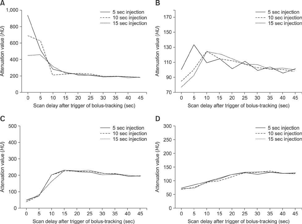
Fig. 2 The change curves of mean attenuation values at the aorta (A), pancreatic parenchyma (B), portal vein (C), and liver parenchyma (D) versus scan delays of 5 sec after automatic triggering by the bolus-tracking technique with the injection durations.
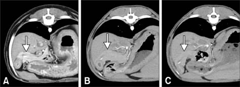
Fig. 3 Transverse CT images of pancreatic body level obtained using 5 sec scan delay after triggering by the bolus-tracking technique with 5 (A), 10 (B), and 15 (C) sec injection durations in a Beagle dog. Note the difference in pancreatic parenchymal contrast enhancement (arrows) by the injection duration of contrast material.
Reference
-
1. Adibi A, Shahbazi A. Automatic bolus tracking versus fixed time-delay technique in biphasic multidetector computed tomography of the abdomen. Iran J Radiol. 2014; 11:e4617.
Article2. Bae KT. Intravenous contrast medium administration and scan timing at CT: considerations and approaches. Radiology. 2010; 256:32–61.
Article3. Bae KT. Peak contrast enhancement in CT and MR angiography: when does it occur and why? pharmacokinetic study in a porcine model. Radiology. 2003; 227:809–816.
Article4. Bae KT. Test-bolus versus bolus-tracking techniques for CT angiographic timing. Radiology. 2005; 236:369–370.
Article5. Bae KT, Heiken JP, Brink JA. Aortic and hepatic contrast medium enhancement at CT. Part II. Effect of reduced cardiac output in a porcine model. Radiology. 1998; 207:657–662.
Article6. Bae KT, Heiken JP, Brink JA. Aortic and hepatic peak enhancement at CT: effect of contrast medium injection rate—pharmacokinetic analysis and experimental porcine model. Radiology. 1998; 206:455–464.
Article7. Bonaldi VM, Bret PM, Atri M, Garcia P, Reinhold C. A comparison of two injection protocols using helical and dynamic acquisitions in CT examinations of the pancreas. AJR Am J Roentgenol. 1996; 167:49–55.
Article8. Cáceres AV, Zwingenberger AL, Hardam E, Lucena JM, Schwarz T. Helical computed tomographic angiography of the normal canine pancreas. Vet Radiol Ultrasound. 2006; 47:270–278.
Article9. Cademartiri F, Nieman K, van der Lugt A, Raaijmakers RH, Mollet N, Pattynama PMT, de Feyter PJ, Krestin GP. Intravenous contrast material administration at 16–detector row helical CT coronary angiography: test bolus versus bolus-tracking technique. Radiology. 2004; 233:817–823.
Article10. Choi SY, Choi HJ, Lee KJ, Lee YW. Establishment of optimal scan delay for multi-phase computed tomography using bolus-tracking technique in canine pancreas. J Vet Med Sci. 2015; 77:1049–1054.
Article11. Claussen CD, Banzer D, Pfretzschner C, Kalender WA, Schörner W. Bolus geometry and dynamics after intravenous contrast medium injection. Radiology. 1984; 153:365–368.
Article12. Fletcher JG, Wiersema MJ, Farrell MA, Fidler JL, Burgart LJ, Koyama T, Johnson CD, Stephens DH, Ward EM, Harmsen WS. Pancreatic malignancy: value of arterial, pancreatic, and hepatic phase imaging with multi–detector row CT. Radiology. 2003; 229:81–90.
Article13. Graf O, Boland GW, Warshaw AL, Fernandez-del-Castillo C, Hahn PF, Mueller PR. Arterial versus portal venous helical CT for revealing pancreatic adenocarcinoma: conspicuity of tumor and critical vascular anatomy. AJR Am J Roentgenol. 1997; 169:119–123.
Article14. Han JK, Choi BI, Kim AY, Kim SJ. Contrast media in abdominal computed tomography: optimization of delivery methods. Korean J Radiol. 2001; 2:28–36.
Article15. Hollett MD, Jorgensen MJ, Jeffrey RB Jr. Quantitative evaluation of pancreatic enhancement during dual-phase helical CT. Radiology. 1995; 195:359–361.
Article16. Holley HC, Koslin DB, Berland LL, Stanley RJ. Inhomogeneous enhancement of liver parenchyma secondary to passive congestion: contrast-enhanced CT. Radiology. 1989; 170:795–800.
Article17. Iseri T, Yamada K, Chijiwa K, Nishimura R, Matsunaga S, Fujiwara R, Sasaki N. Dynamic computed tomography of the pancreas in normal dogs and in a dog with pancreatic insulinoma. Vet Radiol Ultrasound. 2007; 48:328–331.
Article18. Katori R. Normal cardiac output in relation to age and body size. Tohoku J Exp Med. 1979; 128:377–387.
Article19. Kinns J, Malinowski R, McEvoy F, Schwarz T, Zwingenberger A. Special software applications. In : Schwarz T, Saunders J, editors. Veterinary Computed Tomography. West Sussex: Wiley Blackwell;2011. p. 67–74.20. Kondo H, Kanematsu M, Goshima S, Miyoshi T, Shiratori Y, Onozuka M, Moriyama N, Bae KT. MDCT of the pancreas: optimizing scanning delay with a bolus-tracking technique for pancreatic, peripancreatic vascular, and hepatic contrast enhancement. AJR Am J Roentgenol. 2007; 188:751–756.
Article21. Kutara K, Seki M, Ishikawa C, Sakai M, Kagawa Y, Iida G, Ishigaki K, Teshima K, Edamura K, Nakayama T, Asano K. Triple-phase helical computed tomography in dogs with hepatic masses. Vet Radiol Ultrasound. 2014; 55:7–15.
Article22. Mai W, Cáceres AV. Dual-phase computed tomographic angiography in three dogs with pancreatic insulinoma. Vet Radiol Ultrasound. 2008; 49:141–148.
Article23. Nelson NC, Nelson LL. Anatomy of extrahepatic portosystemic shunts in dogs as determined by computed tomography angiography. Vet Radiol Ultrasound. 2011; 52:498–506.
Article24. Platt JF, Reige KA, Ellis JH. Aortic enhancement during abdominal CT angiography: correlation with test injections, flow rates, and patient demographics. AJR Am J Roentgenol. 1999; 172:53–56.
Article25. Puskás Z, Schuierer G. Determination of blood circulation time for optimizing contrast medium administration in CT angiography. Radiologe. 1996; 36:750–757.26. Rivitz SM, Drucker EA. Power injection of peripherally inserted central catheters. J Vasc Interv Radiol. 1997; 8:857–863.
Article27. Rubin GD, Alfrey EJ, Dake MD, Semba CP, Sommer FG, Kuo PC, Dafoe DC, Waskerwitz JA, Bloch DA, Jeffrey RB. Assessment of living renal donors with spiral CT. Radiology. 1995; 195:457–462.
Article28. Shanaman MM, Hartman SK, O'Brien RT. Feasibility for using dual-phase contrast-enhanced multi-detector helical computed tomography to evaluate awake and sedated dogs with acute abdominal signs. Vet Radiol Ultrasound. 2012; 53:605–612.
Article29. Sheiman R, Raptopoulos V, Caruso P, Vrachliotis T, Pearlman J. Comparison of tailored and empiric scan delays for CT angiography of the abdomen. AJR Am J Roentgenol. 1996; 167:725–729.
Article30. Tateishi K, Kishimoto M, Shimizu J, Yamada K. A comparison between injection speed and iodine delivery rate in contrast-enhanced computed tomography (CT) for normal beagles. J Vet Med Sci. 2008; 70:1027–1030.
Article31. Tublin ME, Tessler FN, Cheng SL, Peters TL, McGovern PC. Effect of injection rate of contrast medium on pancreatic and hepatic helical CT. Radiology. 1999; 210:97–101.
Article32. Zwingenberger AL, Schwarz T. Dual-phase CT angiography of the normal canine portal and hepatic vasculature. Vet Radiol Ultrasound. 2004; 45:117–124.
Article33. Zwingenberger AL, Schwarz T, Saunders HM. Helical computed tomographic angiography of canine portosystemic shunts. Vet Radiol Ultrasound. 2005; 46:27–32.
Article
- Full Text Links
-
- Actions
-
Cited
- CITED
-
- Close
- Share
-
- Similar articles
-
- Optimization of scan delay for multi-phase computed tomography by using bolus tracking in normal canine kidney
- Three-dimensional CT angiography of the canine hepatic vasculature
- Computed tomographic anatomy of hepatic artery in normal beagle dogs
- Variation of Attenuation Value of Pancreas at Dual Phase MDCT: Comparison of the Bolus-tracking Technique vs. the Fixed Scan Delay Protocol
- Quantitative Evaluation in Enhancement of Pancreas and Adjacent Vessels during Spiral CT
