A Case of Repeated Dexamethasone Implantation in a Suspected Patient with IRVAN Syndrome
- Affiliations
-
- 1Department of Ophthalmology, Kyung Hee University Hospital, Kyung Hee University School of Medicine, Seoul, Korea. syyu@khu.ac.kr
- KMID: 2362717
- DOI: http://doi.org/10.3341/jkos.2016.57.12.1964
Abstract
- PURPOSE
In the present study, a case of repeated intravitreal dexamethasone implantation for a suspected idiopathic retinal vasculitis, aneurysms and neuroretinitis (IRVAN) syndrome associated with recurrent exudative retinal detachment and macular edema is reported.
CASE SUMMARY
A 39-year-old female who underwent steroid pulse therapy due to Vogt-Koyanagi-Harada disease in the left eye was referred for exudative retinal detachment and macular edema. Best corrected visual acuity (BCVA) was 1.0 in the right eye and 0.5 in the left eye. Cystoid macular edema combined with serous retinal detachment was observed on spectral-domain optical coherence tomography. Fluorescein angiography revealed neovascularization and multiple macroaneurysms with fluorescein leakage in the left peripapillary area. Severe peripheral capillary non-perfusion and fluorescein leakage were also observed in both eyes. Intravitreal dexamethasone implantation was performed in the left eye and macular edema showed wax-and-wane pattern. No edema was observed after 4 additional dexamethasone implantations, however, preretinal hemorrhage occurred in the peripapillary area during treatment. Seventeen months after initiation of treatment, BCVA was 0.6 in the left eye and dry macula was maintained.
CONCLUSIONS
Repeated intravitreal dexamethasone implantation was effective for recurrent macular edema in a patient suspected with IRVAN syndrome.
MeSH Terms
Figure
-
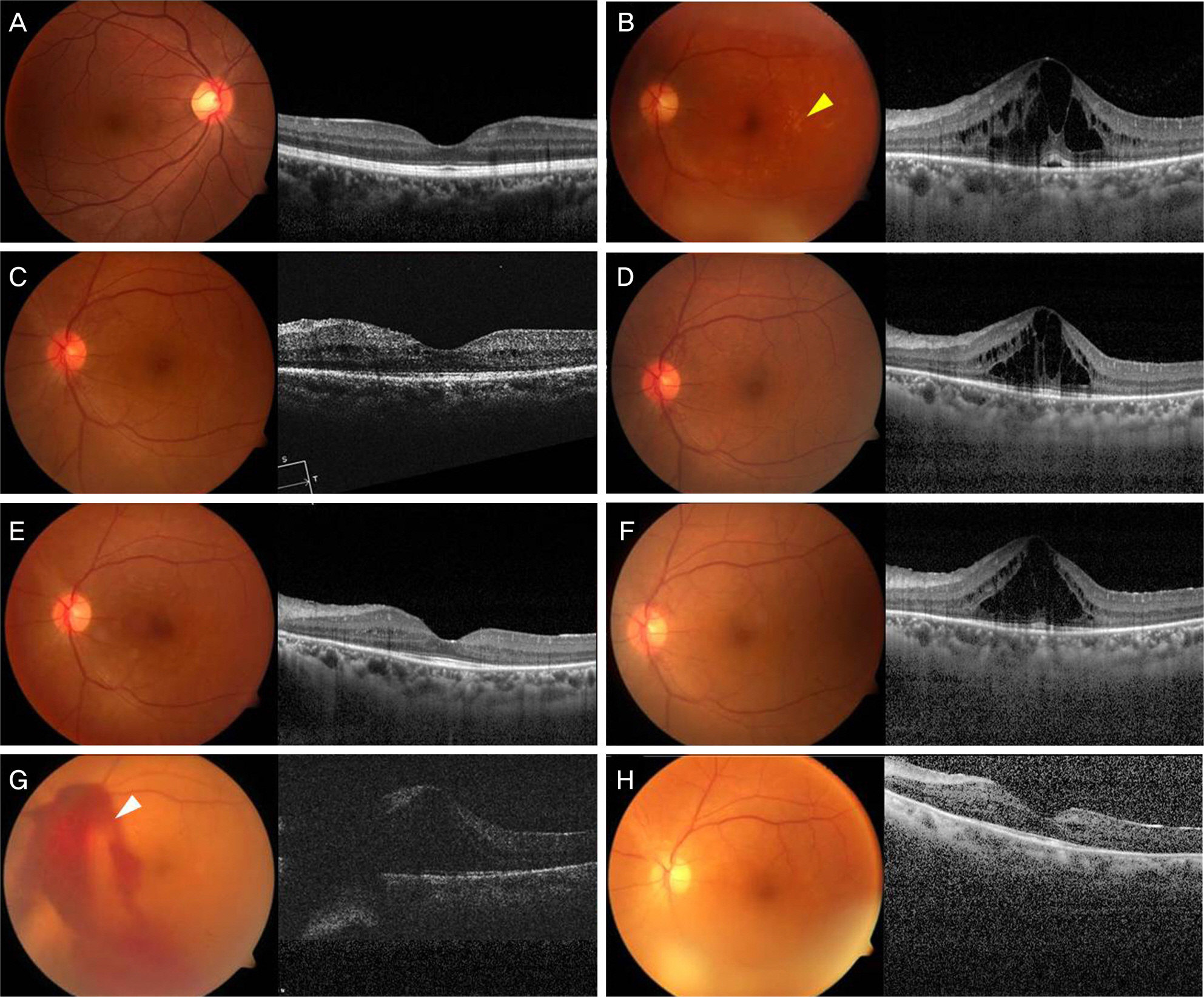
Figure 1. Serial changes of fundus color photographs and optical coherence tomography images of left eye. At the first visit, fundus color photograph showed normal fundus of right eye (A) and aneurysmal dilatations and neovascularization along the retinal arteriolar tree and optic nerve head with intraretinal exudates (yellow arrowhead) on left eye (B). Optical coherence tomography image showed no macular abnormalities on right eye and macular edema combined with serous detachment of retina on left eye. At 2 month after first implantation, macular edema was reduced (C). At 3 months after first implantation, macular edema recurred and second implantation was performed (D). At 3 months after second implantation, complete resolution of macular edema showed and pan-retinal photocoagulation was performed (E). Macular edema recurred at 4 months after second implantation and third implantation was performed (F). At 3 months after third implantation, pre-retinal hemorrhage and macular edema appeared and fourth implantation was performed (white arrowhead) (G). At 2 months after fifth implantation, macular edema got resolved and a moderate cataract was observed (H).
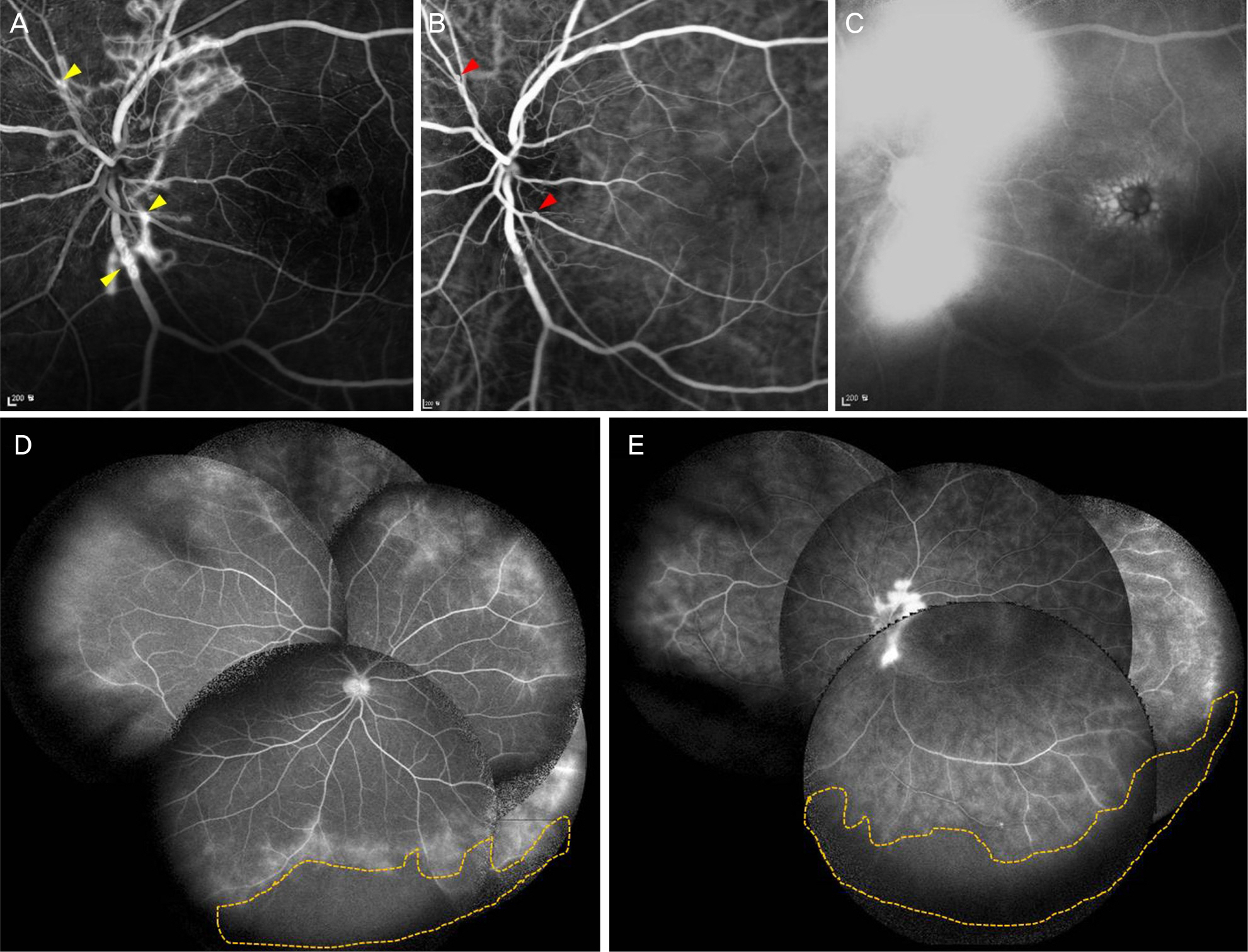
Figure 2. At the first visit. (A) Early fluorescein angiography (FA) reveals neovascularization and macroaneurysms clustered at optic nerve (yellow arrowheads). (B) Indocyanine green angiography of early phase reveals multiple aneurysmal dilatations along the upper temporal and lower nasal retinal arterioles (red arrowheads). (C) FA on late phase demonstrates marked increase of leakage. Wide-field fluorescein angiography reveals peripheral retinal capillary nonperfusion and leakage on right eye (D) and left eye (E) (yellow dot line).
Reference
-
References
1. Chang TS, Aylward GW, Davis JL, et al. Idiopathic retinal abdominal, aneurysms, and abdominal. Retinal Vasculitis Study. Ophthalmology. 1995; 102:1089–97.2. DiLoreto DA Jr, Sadda SR. Idiopathic retinal vasculitis, abdominal, and neuroretinitis (IRVAN) with preserved perfusion. Retina. 2003; 23:554–7.3. Pichi F, Ciardella AP. Imaging in the diagnosis and management of idiopathic retinal vasculitis, aneurysms, and neuroretinitis (IRVAN). Int Ophthalmol Clin. 2012; 52:275–82.
Article4. Samuel MA, Equi RA, Chang TS, et al. Idiopathic retinitis, abdominal, aneurysms, and neuroretinitis (IRVAN): new observations and a proposed staging system. Ophthalmology. 2007; 114:1526–9.e1.5. Tomita M, Matsubara T, Yamada H, et al. Long term follow up in a case of successfully treated idiopathic retinal vasculitis, abdominal, and neuroretinitis (IRVAN). Br J Ophthalmol. 2004; 88:302–3.6. Karagiannis D, Soumplis V, Georgalas I, Kandarakis A. Ranibizumab for idiopathic retinal vasculitis, aneurysms, and abdominal: favorable results. Eur J Ophthalmol. 2010; 20:792–4.7. Empeslidis T, Banerjee S, Vardarinos A, Konstas AG. Dexamethasone intravitreal implant for idiopathic retinal vasculitis, abdominal, and neuroretinitis. Eur J Ophthalmol. 2013; 23:757–60.8. Saatci AO, Ayhan Z, Takeş Ö, et al. Single bilateral dexamethasone implant in addition to panretinal photocoagulation and oral abdominal treatment in IRVAN syndrome. Case Rep Ophthalmol. 2015; 6:56–62.9. Moon SJ, Misch DM. Intravitreal bevacizumab for macular edema from idiopathic retinal vasculitis, aneurysms, and neuroretinitis. Ophthalmic Surg Lasers Imaging. 2010; Mar 9:1–3. doi:. DOI: 10.3928/15428877-20100216-10. [Epub ahead of print].
Article10. Owens SL, Gregor ZJ. Vanishing retinal arterial aneurysms: a case report. Br J Ophthalmol. 1992; 76:637–8.
Article11. Gedik S, Yilmaz G, Akça S, Akova YA. An atypical case of abdominal retinal vasculitis, aneurysms, and neuroretinitis (IRVAN) syndrome. Eye (Lond). 2005; 19:469–71.12. Venkatesh P, Verghese M, Davde M, Garg S. Primary vascular abdominal in IRVAN (idiopathic retinal vasculitis, aneurysms, abdominal) syndrome. Ocul Immunol Inflamm. 2006; 14:195–6.13. Cheema RA, Al-Askar E, Cheema HR. Infliximab therapy for abdominal retinal vasculitis, aneurysm, and neuroretinitis syndrome. J Ocul Pharmacol Ther. 2011; 27:407–10.14. Yeshurun I, Recillas-Gispert C, Navarro-Lopez P, et al. Extensive dynamics in location, shape, and size of aneurysms in a patient with idiopathic retinal vasculitis, aneurysms, and neuroretinitis (IRVAN) syndrome. Idiopathic retinal vasculitis, aneurysms, and neuroretinitis. Am J Ophthalmol. 2003; 135:118–20.15. Sawhney GK, Payne JF, Ray R, et al. Combination anti-VEGF and corticosteroid therapy for idiopathic retinal vasculitis, aneurysms, and neuroretinitis syndrome. Ophthalmic Surg Lasers Imaging Retina. 2013; 44:599–602.
Article
- Full Text Links
-
- Actions
-
Cited
- CITED
-
- Close
- Share
-
- Similar articles
-
- A Case of Repeated Intravitreal Dexamethasone Implantation for Treatment of Macular Edema after Scleral Fixation of Intraocular Lens
- Intraocular Pressure Elevation after 0.7 mg Intravitreal Dexamethasone (Ozurdex(R)) Implantation: A One Year Follow-Up
- Phacoemulsification and Intraocular Lens Implantation after Unexpected Intralenticular Injection of a Dexamethasone Implant
- Intravitreal Dexamethasone Implantation in a Behcet's Disease Patient with Macular Edema, Vasculitis after Cataract Surgery
- A Case of Intravitreal Dexamethasone Implantation in a Patient with Vogt-Koyanagi-Harada Disease
