Spontaneous Osteonecrosis of the Contralateral Medial Tibial Plateau Following Spontaneous Osteonecrosis of Medial Femoral Condyle
- Affiliations
-
- 1Department of Orthopedic Surgery, Kwangju Christian Hospital, Gwangju, Korea. paedic@chol.com
Abstract
- Spontaneous osteonecrosis of the knee (SPONK) is rare disease and most common in the medial femoral condyle. This condition presents with acute onset of pain in elderly patients, usually without a history of trauma. The exact etiology of SPONK is still debated. There are several options for the treatment according to the size, progression and site of the osteonecrosis. SPONK usually occurs in one knee. The spontaneous osteonecrosis of the medial tibial plateau is less recognized than osteonecrosis of the medial femoral condyle. And, in this case, SPONK in the medial tibial plateau of the contralateral knee followed SPONK in the medial femoral condyle, and bony destruction extended to the lateral aspect of the lateral tibial eminence from the medial tibial plateau. The best treatment has not yet been defined. This condition of the tibial side has been managed by total knee replacement resulting in a satisfactory outcome.
Figure
-
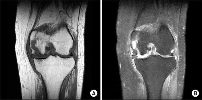
Figure 1 (A) T1 weighted, and (B) T2 weighted coronal magnetic resonance images showed osteonecrosis involving the medial femoral condyle and articular surface of the left knee.

Figure 2 (A) T2 weighted sagittal, and (B) T2 weighted coronal magnetic resonance images taken at the local hospital showed osteonecrosis involving the medial tibial plateau with signal changes extended to the lateral tibial side of the right knee.

Figure 3 (A) Anteroposterior view, and (B) lateral view in our initial plain radiograph at 17 days after local magnetic resonance imaging of the right knee showed a small amount of depression of the medial tibial plateau.

Figure 4 (A, B) Plain radiographs taken at eight months after the initial visit to our hospital showed much progressed destruction and depression of the medial tibial plateau. (C, D) Computed tomography scan images taken on the same day showed bony destruction and osteonecrosis involving the medial tibial condyle and lateral aspect of the intercondylar eminence of the right tibia.

Figure 5 (A) Intraoperative photograph of the right knee showed cartilage irregularity and subchondral bony destruction of the medial tibial condyle and intercondylar eminence. (B) The bony specimen of the medial tibial plateau showed dead bony trabeculae with loss of nuclei and necrotic marrow consistent with avascular necrosis (H&E, ×100).

Figure 6 (A, B) Immediate post-operative radiographs showed that total knee replacement had been performed with metal augmentation for the bony destruction of the medial tibia. (C-E) Plain radiographs taken at six months after the operation showed that artificial devices on the right knee were well positioned but articular defect (arrow) of the left medial femoral condyle was sustained.
Reference
-
1. Ahlbäck S, Bauer GC, Bohne WH. Spontaneous osteonecrosis of the knee. Arthritis Rheum. 1968; 11:705–733.
Article2. Lotke PA, Nelson CL, Lonner JH. Spontaneous osteonecrosis of the knee: tibial plateaus. Orthop Clin North Am. 2004; 35:365–370.
Article3. Jung KA, Lee SC, Hwang SH, Kim DS, Kim TK. Spontaneous osteonecrosis of the knee involving both the medial femoral condyle and the medial tibial plateau: report of three cases. Knee Surg Sports Traumatol Arthrosc. 2008; 16:759–762.
Article4. Soucacos PN, Xenakis TH, Beris AE, Soucacos PK, Georgoulis A. Idiopathic osteonecrosis of the medial femoral condyle. Classification and treatment. Clin Orthop Relat Res. 1997; (341):82–89.
Article5. Schindler OS, Misra R, Spalding TJ. Osteonecrosis of the medial tibial plateau: a case report. J Orthop Surg (Hong Kong). 2006; 14:325–329.
Article6. Lee BI, Choi MK, Rah SK, Choi CU. Spontaneous osteonecrosis of the knee. J Korean Knee Soc. 1994; 6:73–83.7. Lotke PA, Abend JA, Ecker ML. The treatment of osteonecrosis of the medial femoral condyle. Clin Orthop Relat Res. 1982; (171):109–116.
Article8. Kantor H. Bone marrow pressure in osteonecrosis of the femoral condyle (Ahlbäck's disease). Arch Orthop Trauma Surg. 1987; 106:349–352.
Article9. Narváez JA, Narváez J, De Lama E, Sánchez A. Spontaneous osteonecrosis of the knee associated with tibial plateau and femoral condyle insufficiency stress fracture. Eur Radiol. 2003; 13:1843–1848.10. Carpintero P, Leon F, Zafra M, Montero R, Carreto A. Spontaneous collapse of the tibial plateau: radiological staging. Skeletal Radiol. 2005; 34:399–404.
Article
- Full Text Links
-
- Actions
-
Cited
- CITED
-
- Close
- Share
-
- Similar articles
-
- Core Decompression for Post-Arthroscopic Osteonecrosis of the Lateral Tibial Plateau
- Osteonecrosis Occuring in the Medial Femoral Condyle due to Prolonged, Excessive Cortisone Therapy: A Case Report
- Osteonecrosis of the Knee after Arthroscopic Partial Meniscectomy
- Spontaneous Osteonecrosis of the Knee in the Elderly over 60 Years Old
- Autogenous Iliac Tricortical Strut Graft for Bilateral Femoral Bicondylar Osteonecrosis: A Case Report
