Sialadenoma papilliferum: a case report and immunohistochemical study review
- Affiliations
-
- 1Department of Oral and Maxillofacial Surgery, School of Medicine, Institute of Health Sciences, Gyeongsang National University, Jinju, Korea. parkbw@gnu.ac.kr
- 2Department of Pathology, School of Medicine, Gyeongsang National University, Jinju, Korea.
Abstract
- Sialadenoma papilliferum (SP) is a rare benign neoplasm that normally arises from the minor salivary glands, particularly in the palate. SP is normally encountered in older men with an exophytic papillary surface growth. In the present study, an SP of the hard palate of a 69-year-old woman was examined immunohistochemically. Myoepithelial cell markers, such as S-100, smooth muscle actin and vimentin, were observed in the basal or luminal layer of tumor cells, indicating that myoepithelial cells participate in the pathogenesis of SP. In addition, cytokeratin 7 was also strongly detected in the tumor cells, suggesting that excretory ductal epithelial cells have a role in its histogenesis. A review of the literature of immunohistochemical studies on SP showed that the expression and co-expression of cytokeratins and myoepithelial cell markers have been reported in tumor cells. These results suggested that excretory duct cells and myoepithelial cells participate in the pathogenesis of SP.
MeSH Terms
Figure
-

Fig. 1. Perioperative radiologic and clinical features. A. In preoperative MRI view, well demarcated firm mass was observed under the palatal mucosa (white arrow). B. After removal of tumor, intact hard palatal bone was detected. C. Main mass (∗), approximately 1.5x1 cm size, was resected with peripheral normal tissues. D. After 6 months of operation, completely healed palatal mucosa was observed. (MRI: magnetic resonance imaging)
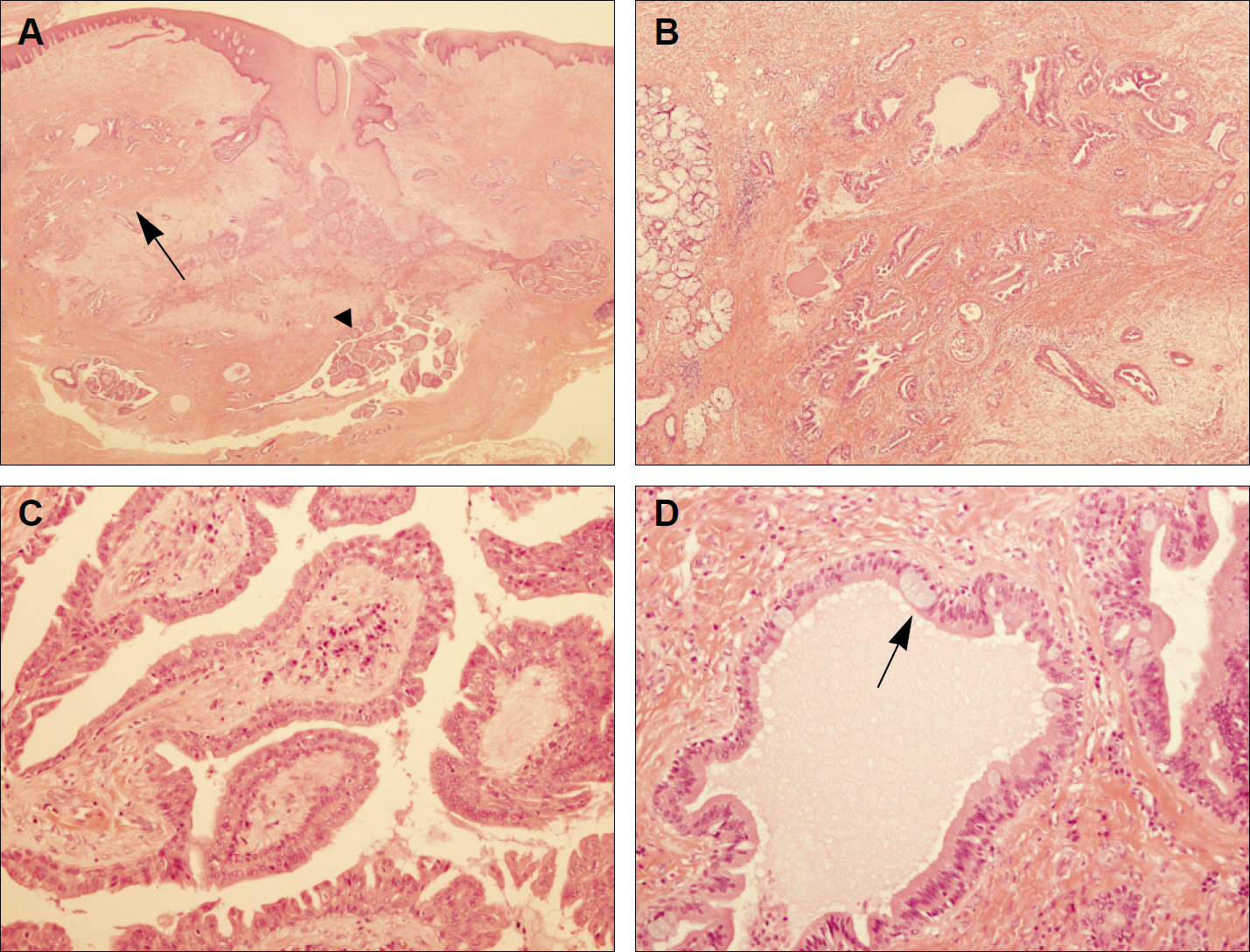
Fig. 2. Photomicrograph of the lesion.(H&E staining) A. The central portion of the tumor was contained hyperplastic squamous epithelium connected to surface mucosa, whereas the remaining tumor showed glandular (arrow) and projecting papillary portion (arrowhead).(original magnification x12) B. Ductal proliferation and dilation with papillary folds were observed in the glandular portion under moderate magnification. (original magnification x40) C. High magnification image of a region showing papillary projection. Papillary fronds were lined by a double row of cells composed of luminal columnar cells and short cuboidal or basaloid cells.(original magnification x200) D. High magnification glandular portion. Some mucin-secreting cells (arrow) were occasionally observed among lining columnar cells of the dilated duct (original magnification x200)
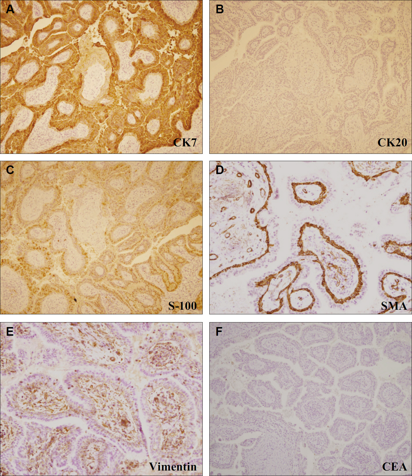
Fig. 3. Immunohistochemical findings of the lesion. A, B. CK 7 was strongly expressed, but CK20 was absent in tumor cells.(original magnification x100) C. S-100 protein was moderately expressed in tumor cell luminal and basal layers.(original magnification x100) D. SMA was strongly expressed in the basal layer of tumor cells (arrow), but was not observed in the luminal columnar cells.(original magnification x200) E. Vimentin was weakly expressed in the basal layer of tumor cells.(original magnification x200) F. CEA expression was not observed in any tumor cell layer. (original magnification x100) (CK7: cytokeratin 7, CK20: cytokeratin 20, S-100: S-100 protein, SMA: smooth muscle actin, CEA: carcinoembryonic antigen)
Reference
-
1. Mahajan D, Khurana N, Setia N. Sialadenoma papilliferum in a young patient: a case report and review of the literature. Oral Surg Oral Med Oral Pathol Oral Radiol Endod. 2007; 103:e51–4.
Article2. Shimoda M, Kameyama K, Morinaga S, Tanaka Y, Hashiguchi K, Shimada M, et al. Malignant transformation of sialadenoma papilliferum of the palate: a case report. Virchows Arch. 2004; 445:641–6.
Article3. Abrams AM, Finck FM. Sialadenoma papilliferum. A previously unreported salivary gland tumor. Cancer. 1969; 24:1057–63.4. Crocker DJ, Christ TF, Cavalaris CJ. Sialadenoma papilliferum: report of case. J Oral Surg. 1972; 30:520–1.5. Markopoulos A, Kayavis I, Papanayotou P. Sialadenoma papilliferum of the oral cavity: report of a case and literature review. J Oral Maxillofac Surg. 1997; 55:1181–4.
Article6. Ubaidat MA, Robinson RA, Belding PJ, Merryman DJ. Sialadenoma papilliferum of the hard palate: report of 2 cases and immunohistochemical evaluation. Arch Pathol Lab Med. 2001; 125:1595–7.7. Fantasia JE, Nocco CE, Lally ET. Ultrastructure of sialadenoma papilliferum. Arch Pathol Lab Med. 1986; 110:523–7.8. Freedman PD, Lumerman H. Sialadenoma papilliferum. Oral Surg Oral Med Oral Pathol. 1978; 45:88–94.
Article9. Maiorano E, Favia G, Ricco R. Sialadenoma papilliferum: an immunohistochemical study of five cases. J Oral Pathol Med. 1996; 25:336–42.
Article10. Nakahata A, Deguchi H, Yanagawa T, Yoshida H, Sato M, Hayashi Y. Coexpression of intermediate-sized filaments in sialadenoma papilliferum and other salivary gland neoplasms. J Oral Pathol Med. 1990; 19:313–8.
Article11. Shirasuna K, Watatani K, Miyazaki T. Ultrastructure of a sialadenoma papilliferum. Cancer. 1984; 53:468–74.
Article12. Bobos M, Hytiroglou P, Karkavelas G, Papakonstantinou C, Papadimitriou CS. Sialadenoma papilliferum of bronchus. Virchows Arch. 2003; 443:695–9.
Article13. Gomes AP, Sobral AP, Loducca SV, de Araújo VC. Sialadenoma papilliferum: immunohistochemical study. Int J Oral Maxillofac Surg. 2004; 33:621–4.
Article14. Rouse RV, Soetikno RM, Baker RJ, Barnard IC, Triadafilopoulos G, Longacre TA. Esophageal submucosal gland duct adenoma. Am J Surg Pathol. 1995; 19:1191–6.
Article15. Pimentel MT, López Amado M, Garcl′a Sarandeses A. Recurrent sialadenoma papilliferum of the buccal mucosa. J Laryngol Otol. 1995; 109:787–90.
Article16. Rennie JS, MacDonald DG, Critchlow HA. Sialadenoma papilliferum. A case report and review of the literature. Int J Oral Surg. 1984; 13:452–4.17. van der Wal JE, van der Waal I. The rare sialadenoma papilliferum. Report of a case and review of the literature. Int J Oral Maxillofac Surg. 1992; 21:104–6.18. Hara K, Ito M, Takeuchi J, Iijima S, Endo T, Hidaka H. Distribution of S-100b protein in normal salivary glands and salivary gland tumors. Virchows Arch A Pathol Anat Histopathol. 1983; 401:237–49.
Article19. de Araujo VC, Carvalho YR, de Araujo NS. Actin versus vimentin in myoepithelial cells of salivary gland tumors. A comparative study. Oral Surg Oral Med Oral Pathol. 1994; 77:387–91.20. Furuse C, Sousa SO, Nunes FD, Magalha ̃es MH, Araújo VC. Myoepithelial cell markers in salivary gland neoplasms. Int J Surg Pathol. 2005; 13:57–65.
Article
- Full Text Links
-
- Actions
-
Cited
- CITED
-
- Close
- Share
-
- Similar articles
-
- A Case of Hidradenoma Papilliferum of the Nipple
- A Case of Ectopic Hidradenoma Papilliferum
- Syringocystadenocarcinoma Papilliferum: A Case Report and Review of the Literature
- A Case of Hidradenoma Papilliferum on the Cheek of an Adult Male
- Hidradenoma Papilliferum of the Anus: A Report of 2 Cases and Review of the Literature
