J Korean Soc Spine Surg.
2001 Mar;8(1):74-80.
Lumbar HIVD Associated with Spondylolysis
- Affiliations
-
- 1Department of Orthopaedic Surgery, Soonchunhyang University, College of Medicine, Bucheon, Korea. schsbj@hosp.sch.ac.kr
Abstract
-
STUDY DESIGN: This is a retrospective study determining the surgical result of lumbar HIVD associated with spondylolysis.
OBJECTIVES
To analyze the incidence of lumbar HIVD associated with spondylolysis and to compare the results of open discectomy for lumbar HIVD associated with spondylolysis to simple lumbar HIVD. SUMMARY OF LITERATURE REVIEW: Lumbar HIVD associated with spondylolysis need be treated by spinal fusion.
MATERIALS AND METHODS
Nine patients(5 males and 4 females) who had lumbar HIVD with spondylolysis, no instability, fol-low-up period of 1yr were identified out of 273 patients with lumbar HIVD, treated by open discectomy from March 1989 to Feb. 1999. The type of HIVD and level of spondylolysis were evaluated, the clinical symptoms and signs including SLR, motor deficit, sensory deficit, change of DTR and severity of radiating pain were periodically followed up on the predesigned protocol.
RESULTS
The incidence of lumbar HIVD associated with spondylolysis is 3.7%. The recovery of back pain was 2.1 to 2.1 by visu-al analogue scale, radiating pain was 7.6 to 0.8. The recovery rate of SLR was 100%, motor deficit; 100%, sensory deficit; 85%, change of DTR; 40%. The clinical evaluation was excellent(2), good(6), fair(1).
CONCLUSIONS
According to the recovery rate of the clinical symptoms, the results of open discectomy for lumbar HIVD associ-ated with spondylolysis without spinal instability and simple HIVD was not different. Therefore, we conclude that lumbar HIVD associated with spondylolysis need not be treated by spinal fusion.
Keyword
Figure
-

Fig. 1-A. Plane radiographs of 49 year-old man with herniated intervertebral disc at L4-5 and L5-S1 and spondylosis at L5 show well demarcated pars interarticularis defect(arrow head). Fig. 1-B. After open discectomy of both L4-5, L5-S1 level, spondylolisthetic anterior translation of L4 vertebral body was not occurred on the last follow-up radiographs.
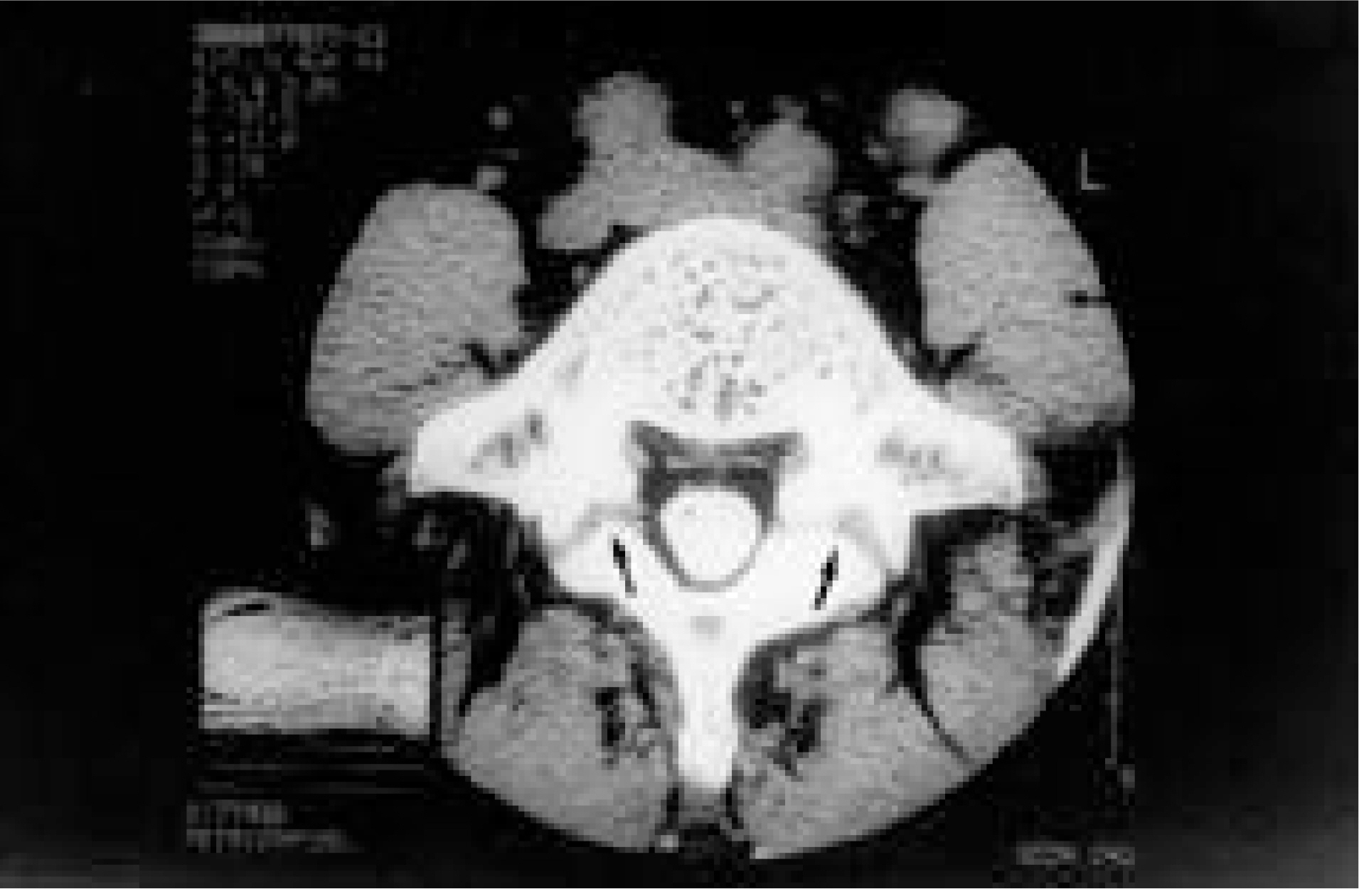
Fig. 2. CT image shows that spondylolytic defects have irregular contours, sclerosis, and loss of cortical bone continuity (arrow head).

Fig. 3-A, B. T2 weighted sagittal and axial MR images show the herniated intervertebral disc at L4-5, L5-S1 level and a low signal intensity area in the left pars interarticularis of L5(arrow head).
Reference
-
1). Alpey AG. System of orthopaedics and fracture, London, Butterworth. 5:224–225. 1978.2). Amato M, Totty WG and Gilula LA. Spondylolysis of the lumbar spine: demonstration of defects and laminal fragmentation. Radiology. 153:627–629. 1984.
Article3). Cauchoix J, Ficat C and Girard B. Repeat surgery after disc excision. Spine. 3:256–259. 1978.
Article4). Fredrickson BE, Baker DR, McHollick WJ, Yuan HA and Lubicky JP. The natural history of spondylolysis and spondylolisthesis. J Bone Joint Surg. 66-A:699–707. 1984.
Article5). Gill K and Frymoyer JW. The management of treatment failures after decompressive surgery. Surgical alternatives and results. In: The Adult Spine Principles and Practice, ed by J Neurosurg. 18:783–789. 1961.6). Gill GG and White HL. Surgical treatment of spondylolisthesis without spine fusion. J Bone Joint Surg. 45-A:666. 1963.7). Grogan JP, Hemminghytt S, Williams AL, Carrera GF and Haughton VM. Spondylolysis studied with computed tomography. Radiology. 145:737–742. 1982.
Article8). Henderson ED. Results of the surgical treatment of spon-dylolisthsis. J Bone Joint Surg. 48-A:619–42. 1966.9). Hensinger RN. Spondylolysis and spondylolisthesis in children and adolescents. J Bone Joint Surg. 71-A:1098–1107. 1989.
Article10). Herron L. Recurrent lumbar disc herniation: Result of repeat laminectomy and discectomy. J of Spinal Disorders. 7:161–166. 1994.11). Iida Y, Kataoka O, Sho T, Sumi M, Hirose T, Bessho Y and Kobayashi D. Postoperative lumbar spinal instability occurring or progressing secondary to laminectomy. Spine. 15:1186–1189. 1990.
Article12). Markwalder M and Battaglia M. Failed back surgery syndrome Part I: Analysis of the clinical presentation and result of testing procedures for instability of the lumbar spine in 171 patients. Acta Neurochirurgica(Wien). 123:46–51. 1993.13). Peter JC, Hoffman EB, Arens LJ and Peacock WJ. Incidence of spinal deformity in children after multiple level laminectomy for selective posterior rhizotomy. Child's Nerv Syst. 6:30–32. 1990.
Article14). Roche MB and Rowe GG. The incidence of separate neural arch and coincident bone variations. J Bone Joint Surg. 34-A:491–494. 1952.15). Rothman SLG and Glenn WV. CT multi-planar recon -struction in 253 cases of lumbar spondylolysis. Am J of Neuroradiol. 5:81–90. 1984.16). Suzuki K, Ishida Y and Ohmori K. Spondylolysis after posterior decompression of the lumbar spine. 35 patients followed for 3-9 years. Acta Orthop Scand. 64:17–21. 1993.
Article17). Tibrewal SB, Pearcy MJ, Portek I and Spivey J. A Prospective study of lumbar spinal movements before and after discectomy using biplanar radiography, correlation of clinical and radiographic findings. Spine. 10:455–460. 1985.18). Tominaga S, Date K and Ohuchi K. Results of extensive laminedtomy for degenerative lumbar canal stenosis. Cent Jpn J Orthop Traumatol. 30:273–275. 1987.19). Wiltse LL. Etiology of spondylolisthesis. Clin Orthop. 10:48–60. 1957.
- Full Text Links
-
- Actions
-
Cited
- CITED
-
- Close
- Share
-
- Similar articles
-
- Radiological Study of Spondylolysis in Korean Laborer
- Herniated Intervertebral Disc of Lumbar Spine in the Teenager
- Segmental Lordosis of the Spondylolytic Vertebrae in Adolescent Lumbar Spondylolysis: Differences between Bilateral L5 and L4 Spondylolysis
- Multidetector CT Findings of Acquired Spondylolysis and Spondylolisthesis after Posterior Lumbar Laminectomy
- Lumbar Spondylolysis and Spondylolytic Spondylolisthesis: Who Should Be Have Surgery? An Algorithmic Approach
