Obstet Gynecol Sci.
2014 Jan;57(1):66-69.
Developed diplopia and ptosis due to a nonfunctioning pituitary macroadenoma during pregnancy
- Affiliations
-
- 1Department of Obstetrics and Gynecology, Hallym University College of Medicine, Seoul, Korea. guittool@hallym.or.kr
Abstract
- Physiologic pituitary enlargement is common during normal pregnancy. However, symptoms such as diplopia, blurred vision and headache resulting from physiologic pituitary enlargement are very rare during pregnancy. A 39-year-old woman complained of sudden diplopia and left eye ptosis at 33th weeks of gestation. An magnetic resonance imaging (MRI) demonstrated the pituitary enlargement compressing the optic chiasm. Notwithstanding the medication of bromocriptine, her symptoms did not regress during pregnancy. At 5 months after delivery, her symptoms dramatically resolved without any surgery, and her visual acuity was normalized. Her MRI scan also revealed more decreased size of pituitary gland compared to antenatal MRI. We report a case of visual loss due to the physiologic pituitary enlargement of nonfunctioning adenoma during pregnancy, which regressed spontaneously after delivery without any surgery.
Keyword
MeSH Terms
Figure
-
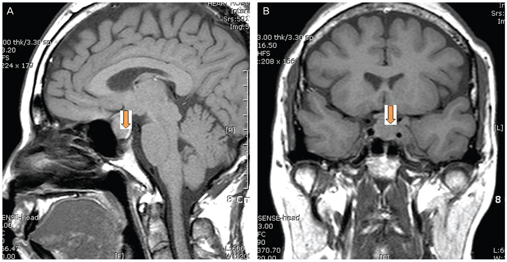
Fig. 1 Pituitary magnetic resonance imaging at pregnancy 34 weeks revealed a large pituitary mass with a height of 1.5 cm and compression of the optic chiasm. Arrows indicate the pituitary mass. (A) Sagittal view. (B) Coronal view.
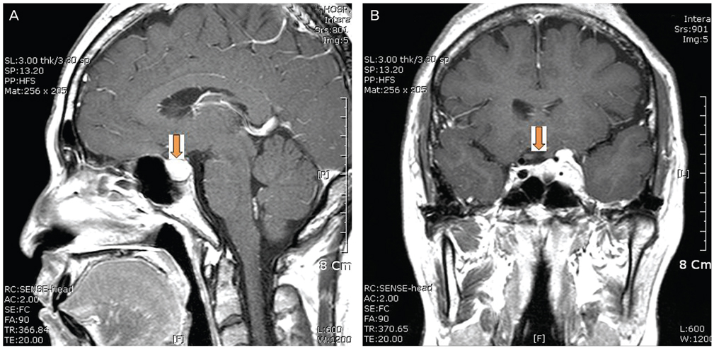
Fig. 2 Pituitary magnetic resonance imaging at postpartum 4 months revealed decreased size (1.3×1.1×0.7 cm) of a pituitary mass. Arrows indicate the pituitary mass. (A) Sagittal view. (B) Coronal view.
Reference
-
1. Dinc H, Esen F, Demirci A, Sari A, Resit Gumele H. Pituitary dimensions and volume measurements in pregnancy and post partum. MR assessment. Acta Radiol. 1998; 39:64–69.2. Molitch ME. Pituitary diseases in pregnancy. Semin Perinatol. 1998; 22:457–470.3. Foyouzi N, Frisbaek Y, Norwitz ER. Pituitary gland and pregnancy. Obstet Gynecol Clin North Am. 2004; 31:873–892.4. Inoue T, Hotta A, Awai M, Tanihara H. Loss of vision due to a physiologic pituitary enlargement during normal pregnancy. Graefes Arch Clin Exp Ophthalmol. 2007; 245:1049–1051.5. Wolpert SM, Molitch ME, Goldman JA, Wood JB. Size, shape, and appearance of the normal female pituitary gland. AJR Am J Roentgenol. 1984; 143:377–381.
- Full Text Links
-
- Actions
-
Cited
- CITED
-
- Close
- Share
-
- Similar articles
-
- A Case of Normal Vaginal Delivery in a Hyperprolactinemia Patient with Pituitary Macroadenoma
- Pituitary Apoplexy due to Pituitary Adenoma Infarction
- A Case of Pituitary Macroadenoma Accompanied with CRH Deficiency
- Comparison of Anterior Pituitary Function between Patients with GH-secreting Macroadenoma and those with Nonfunctioning Macroadenoma
- Clinical Analysis of Pituitary Macroadenoma by Transsphenoidal Approach
