Lab Anim Res.
2010 Sep;26(3):315-318.
Morphometric Analysis of Tibial Bone in Three Strains of Mice Using Micro-computed Tomography
- Affiliations
-
- 1College of Veterinary Medicine, Chonnam National University, Gwangju, Korea. shokim@chonnam.ac.kr
- 2Research Center, Dongnam Institute of Radiological & Medical Sciences (DIRAMS), Busan, Korea.
- 3Advanced Radiation Technology Institute, Jeongeup Campus of Korea Atomic Energy Research Institute, Jeongeup, Korea.
Abstract
- This study investigated the trabecular and cortical bone microarchitecture of tibia in 14-week-old C3H/HeN, C57BL/6J and ICR mice using micro-computed tomography (micro-CT). Defined volumes of interest were scanned at a resolution of 17 micrometer (isotropic). The X-ray tube was set at photon energy of 50 kV, current of 200 microA, exposure time 1.2 sec, and a 0.5 mm-thick aluminium filter. For quantification of bone mineral density (BMD), the bone samples were scanned by micro-CT together with 2 calibration phantoms. The image slices were reconstructed using 3-dimensional CT analyzer software. C3H/HeN mice showed significantly higher levels of bone volume fraction, trabecular number and BMD, and lower levels of trabecular separation, structure model index and degree of anisotropy compared to C57BL/6J or ICR mice in trabecular bone area. So the C3H/HeN mouse appeared to be a good model animal for the study on the changes of trabecular bone with high trabecular bone mass.
Figure
-
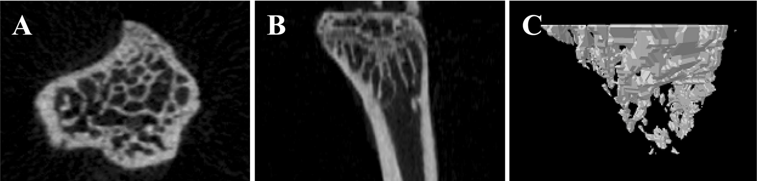
Figure 1. Micro-CT images of the tibia of 14-week-old C3H/HeN mice. Cross sectional view (A), vertical view (B) and reconstructed three-dimensional image (C) are represented.
Reference
-
Bandstra E.R.., Pecaut M.J.., Anderson E.R.., Willey J.S.., De Carlo F.., Stock S.R.., Gridley D.S.., Nelson G.A.., Levine H.G.., Bateman T.A.2008. Longterm dose response of trabecular bone in mice to proton radiation. Radiat. Res. 169(6):607–614.
ArticleBlake G.M.., Fogelman I.2001. Bone densitometry and the diagnosis of osteoporosis. Semin. Nucl. Med. 31(1):69–81.
ArticleBoyd S.K.., Mattmann C.., Kuhn A.., Mller R.., Gasser J.A.2004. A novel approach for monitoring and predicting bone microstructure in osteoporosis. In 26th American Society of Bone and Mineral Research Annual Meeting, Seattle. J. Bone Miner. Res. S236-S237.Eastell R.., Hannon R.A.2008. Biomarkers of bone health and osteoporosis risk. Proc. Nutr. Soc. 67(2):157–162.
ArticleEurell J.A.., Frappier B.L.2006. Textbook of Veterinary Histology, 6th ed., pp. 46–50. Blackwell Publishing, Oxford.Guldberg R.E.., Lin A.S.., Coleman R.., Robertson G.., Duvall C.2004. Microcomputed tomography imaging of skeletal development and growth. Birth Defects Res. C Embryo Today. 72(3):250–259.
ArticleKim S.J.., Kim K.W.., Lee J.H.2000. A study on the trabecular change of femur according to 17β-estradiol dosage in ovariectomized rat. J. Korean Assoc. Maxillofac. Plast. Reconstr. Surg. 22:155–163.Klinck R.J.., Campbell G.M.., Boyd S.K.2008. Radiation effects on bone architecture in mice and rats resulting from in vivo micro-computed tomography scanning. Med. Eng. Phys. 30(7):888–895.
ArticleMayo-Smith W.., Rosenthal D.I.1991. Radiographic appearance of osteopenia. Radiol. Clin. North Am. 29(1):37–47.
- Full Text Links
-
- Actions
-
Cited
- CITED
-
- Close
- Share
-
- Similar articles
-
- Quantification of Microstructures in Mice Alveolar Bone using Micro-computed tomography (microCT)
- Micro-CT analysis of LPS-induced Alveolar Bone Loss in Diabetic Mice
- Study of bony trabecular characteristics using bone morphometry and micro-CT
- The Effects of Antihypertensive Drugs on Bone Mineral Density in Ovariectomized Mice
- Micro-computed tomography analysis of changes in the periodontal ligament and alveolar bone proper induced by occlusal hypofunction of rat molars
