Encapsulated Unresolved Subdural Hematoma Mimicking Acute Epidural Hematoma: A Case Report
- Affiliations
-
- 1Department of Neurosurgery, Presbyterian Medical Center, University of Seonam College of Medicine, Jeonju, Korea. hisarang@hanmail.net
- KMID: 2256254
- DOI: http://doi.org/10.13004/kjnt.2014.10.2.142
Abstract
- Encapsulated acute subdural hematoma (ASDH) has been uncommonly reported. To our knowledge, a few cases of lentiform ASDH have been reported. The mechanism of encapsulated ASDH has been studied but not completely clarified. Encapsulated lentiform ASDH on a computed tomography (CT) scan mimics acute epidural hematoma (AEDH). Misinterpretation of biconvex-shaped ASDH on CT scan as AEDH often occurs and is usually identified by neurosurgical intervention. We report a case of an 85-year-old man presenting with a 2-day history of mental deterioration and right-sided weakness. CT scan revealed a biconvex-shaped hyperdense mass mixed with various densities of blood along the left temporoparietal cerebral convexity, which was misinterpreted as AEDH preoperatively. Emergency craniectomy was performed, but no AEDH was found beneath the skull. In the subdural space, encapsulated ASDH was located. En block resection of encapsulated ASDH was done. Emergency craniectomy confirmed that the preoperatively diagnosed AEDH was an encapsulated ASDH postoperatively. Radiologic studies of AEDH-like SDH allow us to establish an easy differential diagnosis between AEDH and ASDH by distinct features. More histological studies will provide us information on the mechanism underlying encapsulated ASDH.
MeSH Terms
Figure
-
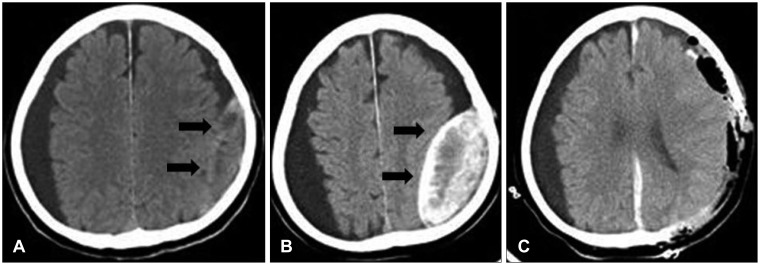
FIGURE 1 A: Incompletely resolved chronic subdural hematoma after burr hole trephination 4 years prior to admission. A thick-walled isodense lesion is seen in the left temporoparietal cerebral convexity. B: Preoperative computed tomography scan shows a lentiform lesion (8×3.8 cm) with high density partially mixed with isodensity to low density, in the left temporoparietal cerebral convexity. Large amounts of subdural fluid collection along both cerebral convexities are seen. C: Postoperative CT scan shows a newly developed hematoma. An acute subdural hematoma along the interhemispheric fissure and left cerebral convexity is seen.
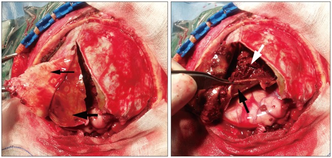
FIGURE 2 Photograph of the encapsulated hematoma in the subdural space shows the dura mater (thin black arrow), outer/inner neomembrane (thick black arrow), solid fresh blood clots with old mud-like blood clots (white arrow), and an intact brain parenchyma.

FIGURE 3 Photograph of resected hematoma. The thick outer neomembrane shows hyaline degeneration, and the thin inner membrane has meningothelial cells and hemosiderin pigments produced by old hemorrhage. A: The dura membrane has plentiful vascular structures (arrow). B: Hemosiderin pigments (arrow). C: Meningothelial cells (arrow). Hematoxylin and eosin staining. Original magnifications are marked by bars.
Reference
-
1. Friede RL. Incidence and distribution of neomembranes of dura mater. J Neurol Neurosurg Psychiatry. 1971; 34:439–446. PMID: 5096558.
Article2. Friede RL, Schachenmayr W. The origin ofsubdural neomembranes. II. Fine structural of neomembranes. Am J Pathol. 1978; 92:69–84. PMID: 686149.3. Kawano N, Endo M, Saito M, Yada K. [Origin of the capsule of a chronic subdural hematoma--an electron microscopy study]. No Shinkei Geka. 1988; 16:747–752. PMID: 3412561.4. Killeffer JA, Killeffer FA, Schochet SS. The outer neomembrane of chronic subdural hematoma. Neurosurg Clin N Am. 2000; 11:407–412. PMID: 10918009.
Article5. Lee KS. Natural history of chronic subdural haematoma. Brain Inj. 2004; 18:351–358. PMID: 14742149.6. Miki S, Fujita K, Katayama W, Sato M, Kamezaki T, Matsumura A, et al. Encapsulated acute subdural hematoma mimicking acute epidural hematoma on computed tomography. Neurol Med Chir (Tokyo). 2012; 52:826–828. PMID: 23183078.
Article7. Oh HJ, Lee KS, Shim JJ, Yoon SM, Yun IG, Bae HG. Postoperative course and recurrence of chronic subdural hematoma. J Korean Neurosurg Soc. 2010; 48:518–523. PMID: 21430978.
Article8. Park HR, Lee KS, Shim JJ, Yoon SM, Bae HG, Doh JW. Multiple Densities of the Chronic Subdural Hematoma in CT Scans. J Korean Neurosurg Soc. 2013; 54:38–41. PMID: 24044079.
Article9. Prieto R, Pascual JM, Subhi-Issa I, Yus M. Acute epidural-like appearance of an encapsulated solid non-organized chronic subdural hematoma. Neurol Med Chir (Tokyo). 2010; 50:990–994. PMID: 21123983.10. Su IC, Wang KC, Huang SH, Li CH, Kuo LT, Lee JE, et al. Differential CT features of acute lentiform subdural hematoma and epidural hematoma. Clin Neurol Neurosurg. 2010; 112:552–556. PMID: 20483531.
Article11. Yamashima T, Yamamoto S. How do vessels proliferate in the capsule of a chronic subdural hematoma? Neurosurgery. 1984; 15:672–678. PMID: 6209593.
Article12. Yatsuzuka H. [Growing factors of chronic subdural hematoma--significance of CK activity in hematoma contents and neomembrane]. No To Shinkei. 1988; 40:963–969. PMID: 3196500.
- Full Text Links
-
- Actions
-
Cited
- CITED
-
- Close
- Share
-
- Similar articles
-
- Intraoperative Development of Contralateral Subdural Hematoma during Evacuation of Acute Subdural Hematoma: Case Report
- Postoperative Contralateral Supra- and Infratentorial Acute Epidural Hematoma after Decompressive Surgery for an Acute Subdural Hematoma: A Case Report
- Bilateral Acute Subdural Hematoma Following Evacuation of Chronic Subdural Hematoma
- Spontaneously Rapid Resolution of Acute Subdural Hemorrhage with Severe Midline Shift
- Acute Cervical Subdural Hematoma with Quadriparesis after Cervical Transforaminal Epidural Block
