Korean J Hematol.
2009 Dec;44(4):320-324. 10.5045/kjh.2009.44.4.320.
Generalized Primary Amyloid Lymphadenopathy
- Affiliations
-
- 1Cancer Research Institute, Seoul National University College of Medicine, Seoul, Korea. ssysmc@snu.ac.kr
- 2Department of Internal Medicine, Seoul National University College of Medicine, Seoul, Korea.
- 3Clinical Research Institute, Seoul National University Hospital, Seoul, Korea.
- 4Department of Pathology, Seoul National University College of Medicine, Seoul, Korea.
- KMID: 2252120
- DOI: http://doi.org/10.5045/kjh.2009.44.4.320
Abstract
- Systemic amyloidosis is a disease that displays deposition of insoluble polymeric protein fibrils in tissues and organs. We report here on a case of a 64-year-old woman who initially presented with multiple enlarged lymph nodes. Computed tomography showed multiple enlarged lymph nodes in the mediastinal, lower cervical, supraclavicular, axillary and abdominal areas. Excision biopsy of the cervical lymph nodes and the subsequent histopathology showed amorphous eosinophilic material deposits, and these revealed apple-green birefringence on a polarizing microscopic examination on the Congo-red stained slide. The patient was diagnosed with amyloidosis and she received chemotherapy consisting of melphalan and dexamethasone. During chemotherapy, she was diagnosed with breast cancer. After modified unilateral radical mastectomy, the dexamethasone was restarted and this therapy resulted in stable disease.
MeSH Terms
Figure
-
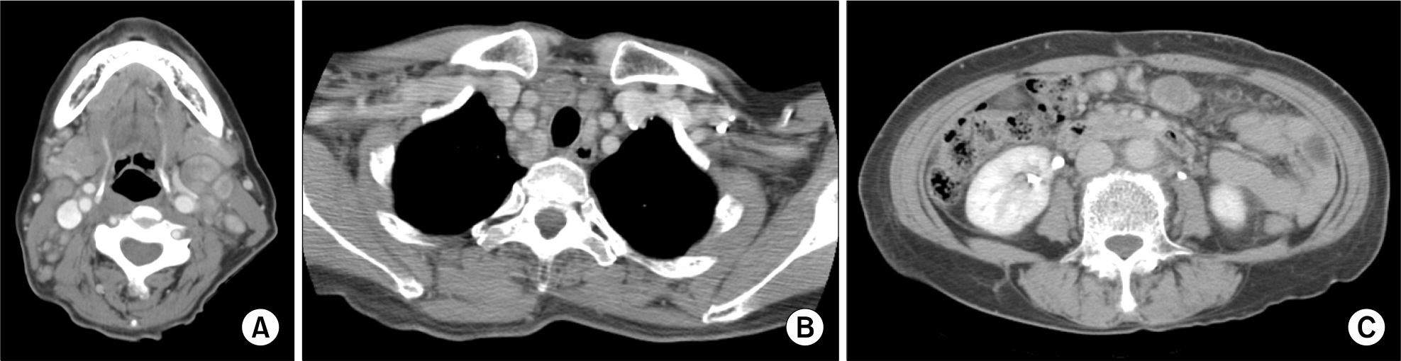
Fig. 1. Computerized tomography (CT) scans at diagnosis showing multiple lymph node enlargements with high enhancement in the cervical (A), mediastinal (B), and abdominal (C) area.
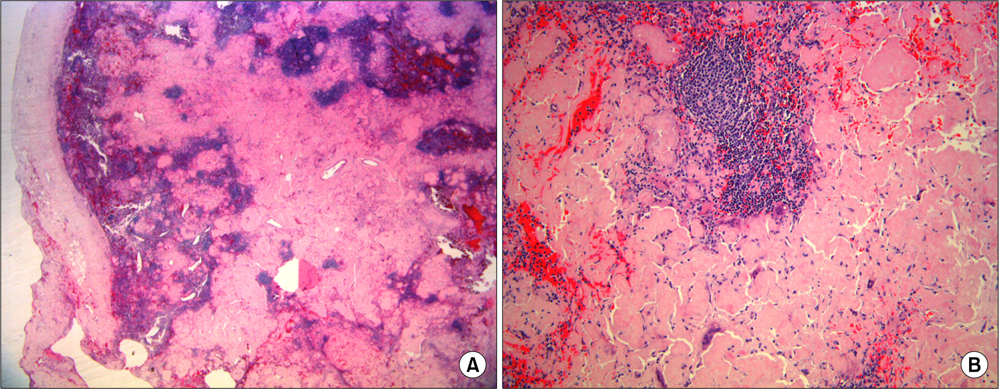
Fig. 2. Cervical lymph node biopsy specimens: H & E stain (×10) (A), and H & E stain (×100) (B) showing amorphous eosinophilic material deposits and chronic inflammation with multinucleated giant cells.
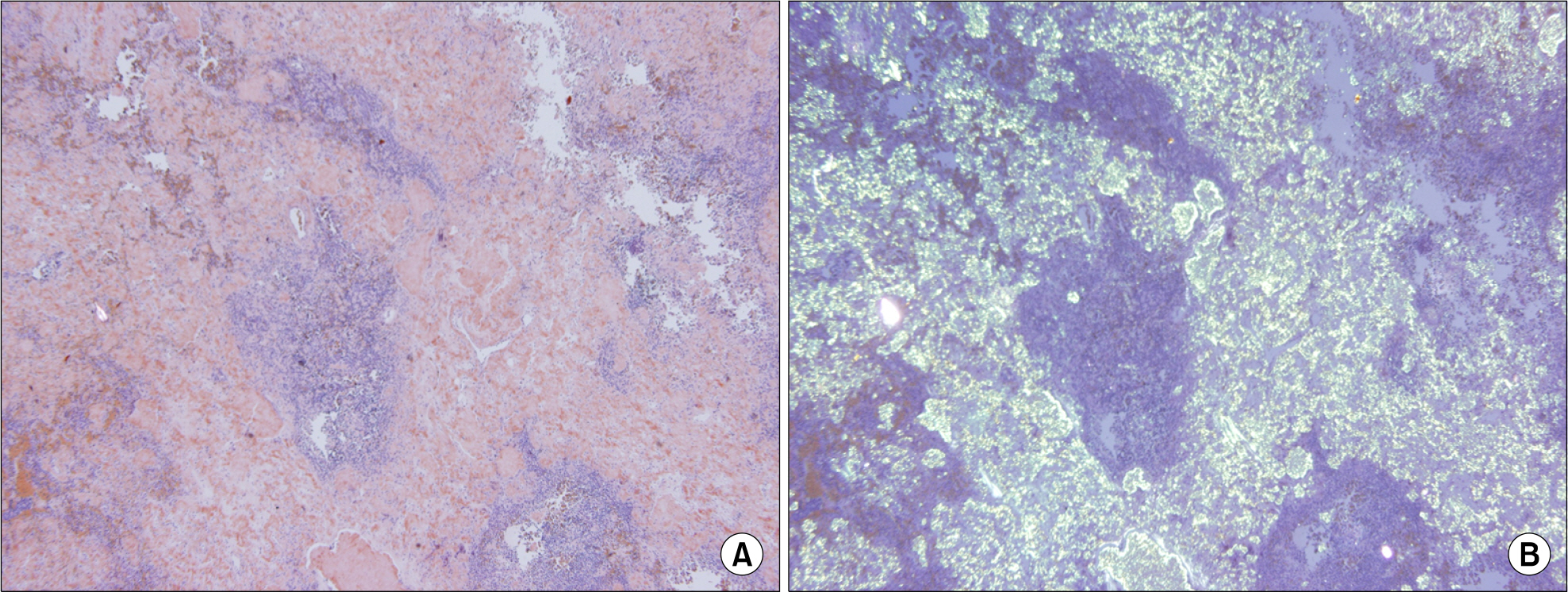
Fig. 3. Congo red stain (×40) of cervical lymph node: Amorphous eosinophilic material deposit (A) and apple-green birefringence (B).
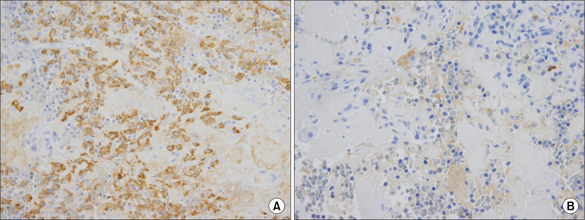
Fig. 4. Immunohistochemical stain for lambda light chain was positive (A), but negative for kappa light chain (B). (Polymer method, ×400).
Reference
-
References
1. Westermark P, Benson MD, Buxbaum JN, et al. Amyloid: toward terminology clarification. Report from the nomenclature committee of the international society of amyloidosis. Amyloid. 2005; 12:1–4.
Article2. Matsuda M, Gono T, Shimojima Y, et al. AL amyloidosis manifesting as systemic lymphadenopathy. Amyloid. 2008; 15:117–24.
Article3. Wechalekar AD, Hawkins PN, Gillmore JD. Perspectives in treatment of AL amyloidosis. Br J Haematol. 2008; 140:365–77.
Article4. Leach DB, Hester TO, Farrell HA, Chowdhury K. Primary amyloidosis presenting as massive cervical lymphadenopathy with severe dyspnea: a case report and review of the literature. Otolaryngol Head Neck Surg. 1999; 120:560–4.5. Ko HS, Davidson JW, Pruzanski W. Amyloid lymphadenopathy. Ann Intern Med. 1976; 85:763–4.
Article6. Rajkumar SV, Gertz MA. Advances in the treatment of amyloidosis. N Engl J Med. 2007; 356:2413–5.
Article7. Skinner M, Sanchorawala V, Seldin DC, et al. High-dose melphalan and autologous stem-cell transplantation in patients with AL amyloidosis: an 8-year study. Ann Intern Med. 2004; 140:85–93.
Article8. Comenzo RL, Gertz MA. Autologous stem cell transplantation for primary systemic amyloidosis. Blood. 2002; 99:4276–82.
Article9. Vogel MN, Wehrmann M, Horger MS. Massive cervical and abdominal lymphadenopathy caused by localized amyloidosis. J Clin Oncol. 2007; 25:343–4.
Article10. Sanchorawala V, Wright DG, Seldin DC, et al. An overview of the use of high-dose melphalan with autologous stem cell transplantation for the treatment of AL amyloidosis. Bone Marrow Transplant. 2001; 28:637–42.
Article
- Full Text Links
-
- Actions
-
Cited
- CITED
-
- Close
- Share
-
- Similar articles
-
- Primary Tracheobronchial Amyloidosis: A Case Report
- A Case of Cerebral Amyloid Angiopathy-related Intracerebral Hemorrhage
- A Case of the Primary Amyloid Polyneuropathy
- Toxocariasis Mimicking Lymphoma and Presenting as Multiple Lymphadenopathy: A Case Report
- Primary Amyloidosis Involving Mediastinal and Cervical Lymph Nodes: A Case Report
