A Giant Unruptured Aneurysm of Distal Internal Carotid Artery Presenting with Compressive Optic Neuropathy
- Affiliations
-
- 1Department of Ophthalmology, Dongkang Medical Center, Ulsan, Korea. nkkoo77@naver.com
- KMID: 2216095
- DOI: http://doi.org/10.3341/jkos.2012.53.9.1368
Abstract
- PURPOSE
To report a case of compressive optic neuropathy due to a giant unruptured aneurysm of a distal internal carotid artery.
CASE SUMMARY
A 68-year-old female presented with a one-week history of visual disturbance in her left eye. The patient had no underlying disease except hypertension. Best corrected visual acuity was 20/20 in the right eye and 8/20 in the left eye. The color perception test showed abnormal findings in the left eye. Slit lamp examination showed no abnormal finding except incipient cataract in both eyes. Additionally, fundus examination showed no abnormal finding. Brain MRI and MRA revealed a 2.4 x 2.2 x 3.0-cm-sized unruptured giant aneurysm on the left internal carotid artery.
CONCLUSIONS
A giant aneurysm should be considered as a cause for acute or subacute optic neuropathy in a patient with hypertension.
MeSH Terms
Figure
-

Figure 1 Fundus photographs show no abnormal findings in both eyes.
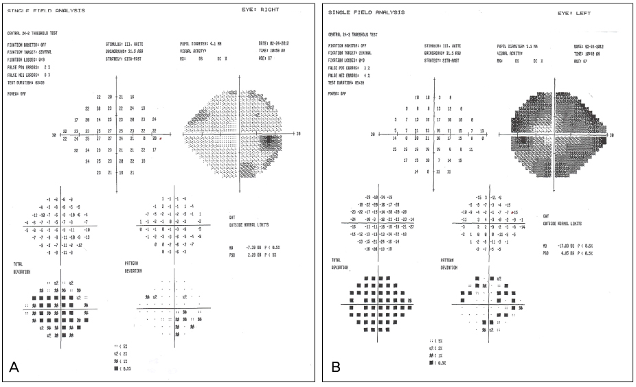
Figure 2 Humphrey visual field test of both eyes. (A) Right eye shows inferotemporal visual field defect on pattern deviation. (B) Left eye shows constricted peripheral visual field on pattern deviation.

Figure 3 MR image of both horizontal section and coronal section. (A) MR images shows abnormal signal on horizontal section (arrow). (B) MR images shows abnormal signal compressing the optic chiasm (arrow) on coronal section.

Figure 4 MR angiogram showing 2.4 × 2.2 × 3.0 cm sized giant intracranial aneurysm of left internal carotid artery (arrow).
Cited by 2 articles
-
Internal Carotid Artery Aneurysm Presenting with Unilateral Nasal Hemianopsia
Kyoung Nam Kim, Chang Sik Kim, Yeon Hee Lee, Sung Bok Lee
J Korean Ophthalmol Soc. 2014;55(9):1406-1411. doi: 10.3341/jkos.2014.55.9.1406.Regrowth of Internal Carotid Artery Aneurysm after Neck Clipping Surgery Presenting with Compressive Optic Neuropathy
Young Nam Kwon, Hak Young Rhee, Yu Jin Jung, Hye-Yeon Choi, Sang-Beom Kim, Won-Chul Shin
Korean J Clin Neurophysiol. 2014;16(2):89-91. doi: 10.14253/kjcn.2014.16.2.89.
Reference
-
1. Artmann H, Vonofakos D, Müller H, Grau H. Neuroradiologic and neuropathologic findings with growing giant intracranial aneurysm. Review of the literature. Surg Neurol. 1984. 21:391–401.2. Horowitz MB, Yonas H, Jungreis C, Hung TK. Management of a giant middle cerebral artery fusiform serpentine aneurysm with distal clip application and retrograde thrombosis: case report and review of the literature. Surg Neurol. 1994. 41:221–225.3. Sonntag VK, Yuan RH, Stein BM. Giant intracranial aneurysms: a review of 13 cases. Surg Neurol. 1977. 8:81–84.4. Kim JY, Choi HY. A case of the giant aneurysm in the distal portion of the posterior cerebral artery: A case report. J Korean Neurosurg Soc. 2000. 29:963–967.5. Sundt TM Jr, Piepgras DG. Surgical approach to giant intracranial aneurysms. Operative experience with 80 cases. J Neurosurg. 1979. 51:731–742.6. Ellamushi H, Thorne L, Kitchen N. Unruptured cerebral aneurysms causing seizure disorder (report of two cases). Seizure. 1999. 8:310–312.7. Heros RC, Kolluri S. Giant intracranial aneurysms presenting with massive cerebral edema. Neurosurgery. 1984. 15:572–577.8. Whittle IR, Dorsch NW, Besser M. Giant intracranial aneurysms: diagnosis, management, and outcome. Surg Neurol. 1984. 21:218–230.9. Fried LC, Yballe A. Rapid formation of giant aneurysms: case report. J Neurol Neurosurg Psychiatry. 1972. 35:527–530.10. Koshikawa N, Kamio M, Sekino H, et al. [Giant aneurysm--a case report with review of literature (author's transl)]. No Shinkei Geka. 1980. 8:79–88.
- Full Text Links
-
- Actions
-
Cited
- CITED
-
- Close
- Share
-
- Similar articles
-
- Regrowth of Internal Carotid Artery Aneurysm after Neck Clipping Surgery Presenting with Compressive Optic Neuropathy
- Compressive Optic Neuropathy Caused by Internal Carotid Artery Aneurysm
- Monocular Superior Altitudinal Field defect due to Supraclinoid Internal Carotid Artery Aneurysm
- Compressive Optic Neuropathy Caused by Internal Carotid Artery Aneurysm Presenting with Concurrent Neuromyelitis Optica
- A Case of Intracavernous Carotid Aneurysm Presenting with Visual Loss with No Oculomotor Disturbance
