J Korean Ophthalmol Soc.
2010 Nov;51(11):1532-1536. 10.3341/jkos.2010.51.11.1532.
A Case of Bilateral Morning Glory Syndrome
- Affiliations
-
- 1Department of Ophthalmology, Kim's Eye Hospital, Myung-Gok Eye Research Institute Konyang University, Seoul, Korea. han66139@kimeye.com
- KMID: 2213992
- DOI: http://doi.org/10.3341/jkos.2010.51.11.1532
Abstract
- PURPOSE
To report a bilateral case of morning glory syndrome.
CASE SUMMARY
On May 30, 2002, a six-year-old patient visited our clinic with impaired visual acuity of her left eye and was diagnosed as having a cataract on her left eye superimposed on a bilateral morning glory anomaly. According to cycloplegic refraction, the patient's corrected visions was 0.8 in the right eye and 0.1 in the left. On June 20, 2002, the patient received ultrasonographic phacoemulsification, intraocular lens implantation, synechiolysis and partial vitectomy in her left eye. Approximately 5.2 years after the surgery, according to manifest refraction, the patient's corrected visions were 0.8 in the right eye and 0.1 in the left, no prominent postoperative changes were observed on slit lamp microscopy and fundus examinations. The thickness of the central macula of her right eye had decreased according to optical coherence tomography; the physiologic scotoma size of the patient's right eye had increased with narrowed peripheral visual field of her left eye.
CONCLUSIONS
Although monocular morning glory anomaly has previously been reported to occur, in the present study case, the anomaly occurred bilaterally. In morning glory patients, strabismus examination and additional evaluation of a patient's general state should be performed along with a regular fundus examination.
MeSH Terms
Figure
-
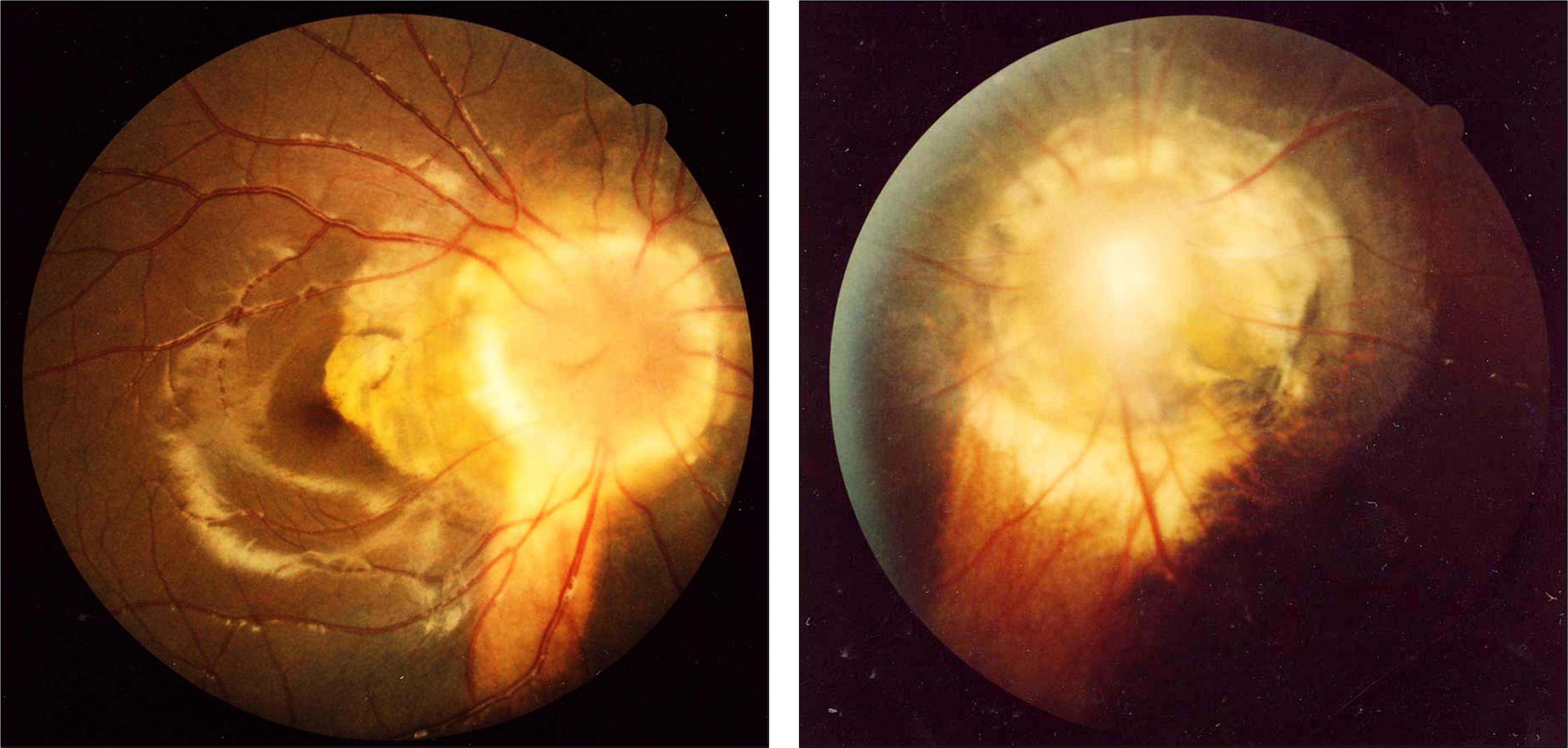
Figure 1. Fundus photographs of the patient with a funnel-shaped excavation.

Figure 2. Disc photographs of the patient shows the blood vessels emerging from the rim of the excavation in a radial pattern like the spokes of a wheel.

Figure 3. Optical coherence tomography shows decreased central macular thickness of right eye (central macula thickness, A = 74 μm).
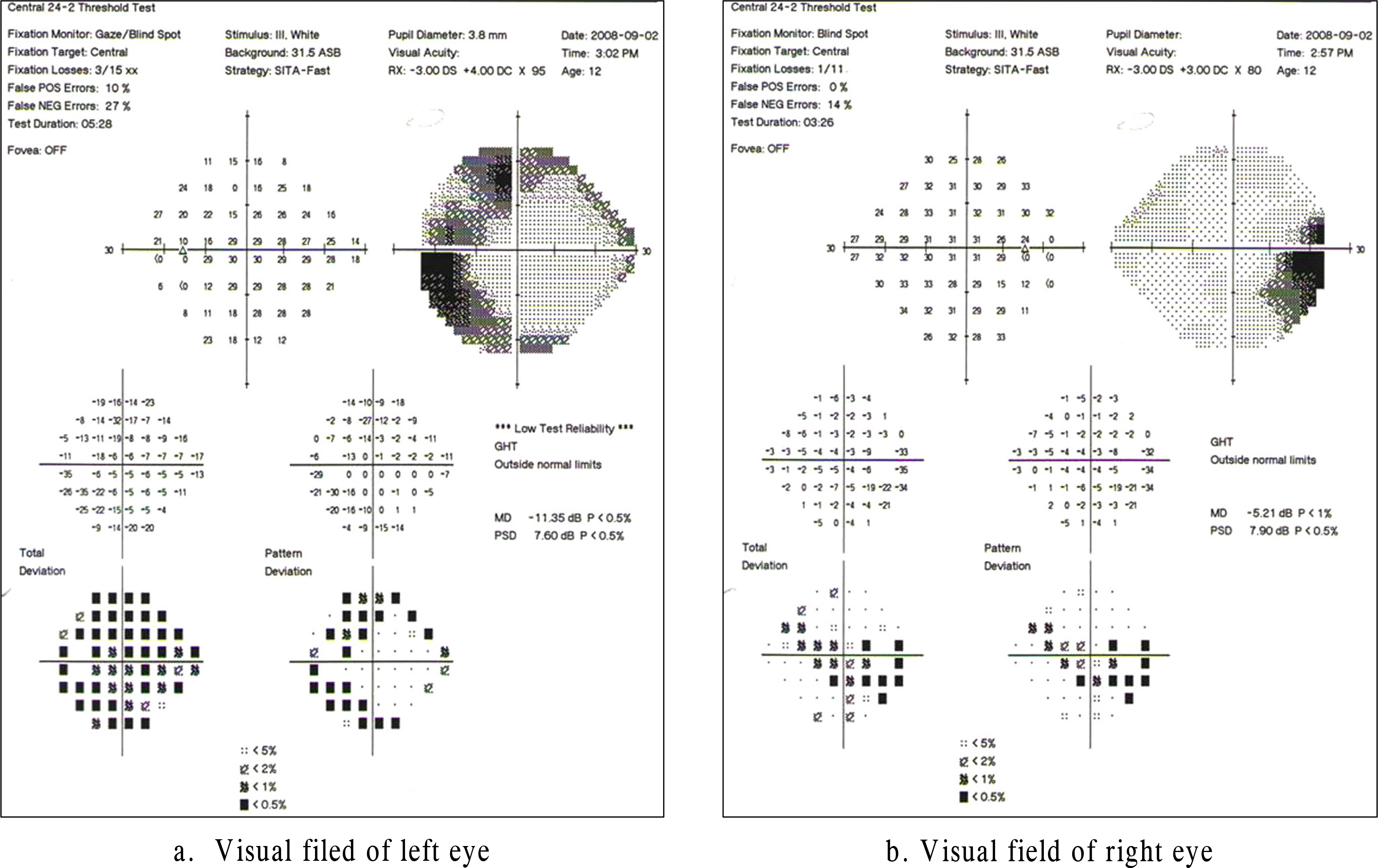
Figure 4. Visual field test (Kinetic and 24–2) shows decreased peripheral visual field of the left eye and increased physiologic scotoma of the right eye.
Reference
-
References
1. Kindler P. Morning glory syndrome: unusual congenital optic disk anomaly. Am J Ophthalmol. 1970; 69:376–84.
Article2. Krause U. Three cases of the morning glory syndrome. Acta Ophthalmol (Copenh). 1972; 50:188–98.
Article3. Steinkuller PG. The morning glory disk anomaly: case report and literature review. J Pediatr Ophthalmol Strabismus. 1980; 17:81–7.
Article4. Beyer WB, Quencer RM, Osher RH. Morning Glory Syndrome: A functional analysis including fluoresein angiography, aberrations and computerized tomography. Ophthalmology. 1982; 89:1362–7.5. Dempster AG, LEE WR, Forrester JV, McCreath GT. The morning golry syndrome-a mesodermal defect? Ophthalmologica. 1983; 187:222–30.6. Goldhammer Y, Smith JL. Optic nerve anomalies in basal encephalocele. Arch Ophthalmol. 1975; 93:115–8.
Article7. Brodsky MC. Congenital anomalies of the optic disc. Miller NR, Newman NG, editors. Walsh & Hoyt's Clinical Neuro-ophthalmology. 5th ed.Chapter 18. Williams & Wilkins;1998. p. 775–823.8. Itakura T, Miyamoto K, Uematsu Y. Bilateral morning glory syndrome associated with sphenoid encephalocele. Case report. J Neurosurg. 1992; 77:949–51.9. De Laey JJ, Ryckaert S, Leys A. The ‘morning glory' syndrome. Ophthalmic Paediatr Genet. 1985; 5:117–24.
Article10. Akiyama K, Azuma N, Hida T, Uremura Y. Retinal detachment in Morning Glory syndrome. Ophthalmic surg. 1984; 15:841–3.11. Miller NR, Newman NJ, editors. Anormalies of the optic disc. The essentials: Walsh & Hoyl's Clinical Neurophthalmology. 5th ed.Williams & Williams;1999. p. 117–23.12. Deb N, Das R, Roy IS. Bilateral Morning Glory Disc Anomaly. Indian J Ophthalmol. 2003; 51:182–4.13. Nagy V, Kettesy B, Toth K, et al. Morning glory aberrations clinical study of two cases. Klin Monatsbl Augenheilkd. 2002; 219:801–5.14. Pollock S. The morning glory disc anomaly: contractile movement, classification and embryogenesis. Doc Ophthalmol. 1987; 65:439–60.
Article15. Loudot C, Fogliarini C, Baeteman C, et al. Rehabilitation on functional ambylopia in morning glory syndrome. J Fr Ophtalmol. 2007; 30:998–1001.16. Caprioli J, Lesser RL. Basal encephalocele and morning glory syndrome. Br J Ophthalmol. 1983; 67:349–51.
Article17. Apple DJ, Rabb MF, Walsh PM. Congenital anomalies of the optic disc. Surv Ophthalmol. 1982; 27:3.
Article18. Traboulsi EI, P'Neil JF. The spectrum in the morphology of the so-called “morning glory disc anomaly”. J Pediatr Ophthalmol Strabismus. 1988; 25:93–8.
Article19. Yoo YS, Chun CH, Lee JH. A case of morning glory syndrome. J Korean Ophthalmol Soc. 1988; 29:749–52.20. Haik BG, Greenstein SH, Smith ME, et al. Retinal detachment in the morning glory anomaly. Ophthalmology. 1984; 91:1638–47.
Article21. Kawano K, Fugita S. Duane's retraction syndrome associated with morning glory syndrome. J Pediatr Ophthalmol Strabismus. 1981; 18:51–4.
Article22. Savell J, Cook JR. Optic nerve colobomas of autosomal dominant hereditary. Arch Ophthalmol. 1976; 94:395–400.
