J Korean Ophthalmol Soc.
2010 Jul;51(7):1036-1038. 10.3341/jkos.2010.51.7.1036.
A Case of Hydranencephaly With Exotropia
- Affiliations
-
- 1Department of Ophthalmology, Seoul National University College of Medicine, Seoul, Korea. hjm@snu.ac.kr
- 2Department of Ophthalmology, Seoul National University Bundang Hospital, Seongnam, Korea.
- 3Department of Radiology, Seoul National University Bundang Hospital, Seongnam, Korea.
- KMID: 2213799
- DOI: http://doi.org/10.3341/jkos.2010.51.7.1036
Abstract
- PURPOSE
To report a large-angle exotropia, limited adduction, epiblepharon, high myopia and no pupillary light reflex in a patient with hydranencephaly.
CASE SUMMARY
A ten-year-old girl with mental retardation presented with exotropia. The patient could fix only with the right eye and was unable to follow with either eye. The Krimsky test revealed 95 prism diopters of exotropia, and adduction was severely limited in both eyes. Pupillary light reflex was absent in both eyes. Cycloplegic refraction showed high myopia in both eyes. Slit lamp examination revealed lower lid epiblepharon and inferior corneal opacity in the right eye. No abnormal findings in the fundus examination were detected. A computed tomogram of the brain showed that the cerebral hemispheres were replaced by a cystic space filled with cerebrospinal fluid, compatible with hydranencephaly. Recession of the lateral rectus muscle and resection of the medial rectus muscle with epiblepharon repair of the lower lid were performed in both eyes. One week postoperatively, the epiblepharon was corrected, and the Krimsky test showed 16 prism diopters of left intermittent exotropia at near.
CONCLUSIONS
When a combined manifestation of mental retardation, limited adduction, no pupillary light reflex and a large-angle exotropia is present, the possibility of a congenital developmental anomaly of the central nervous system including hydranencephaly should be suspected.
Keyword
MeSH Terms
Figure
-

Figure 1. Gaze photographs show a large angle of exotropia and limited adduction in both eyes.
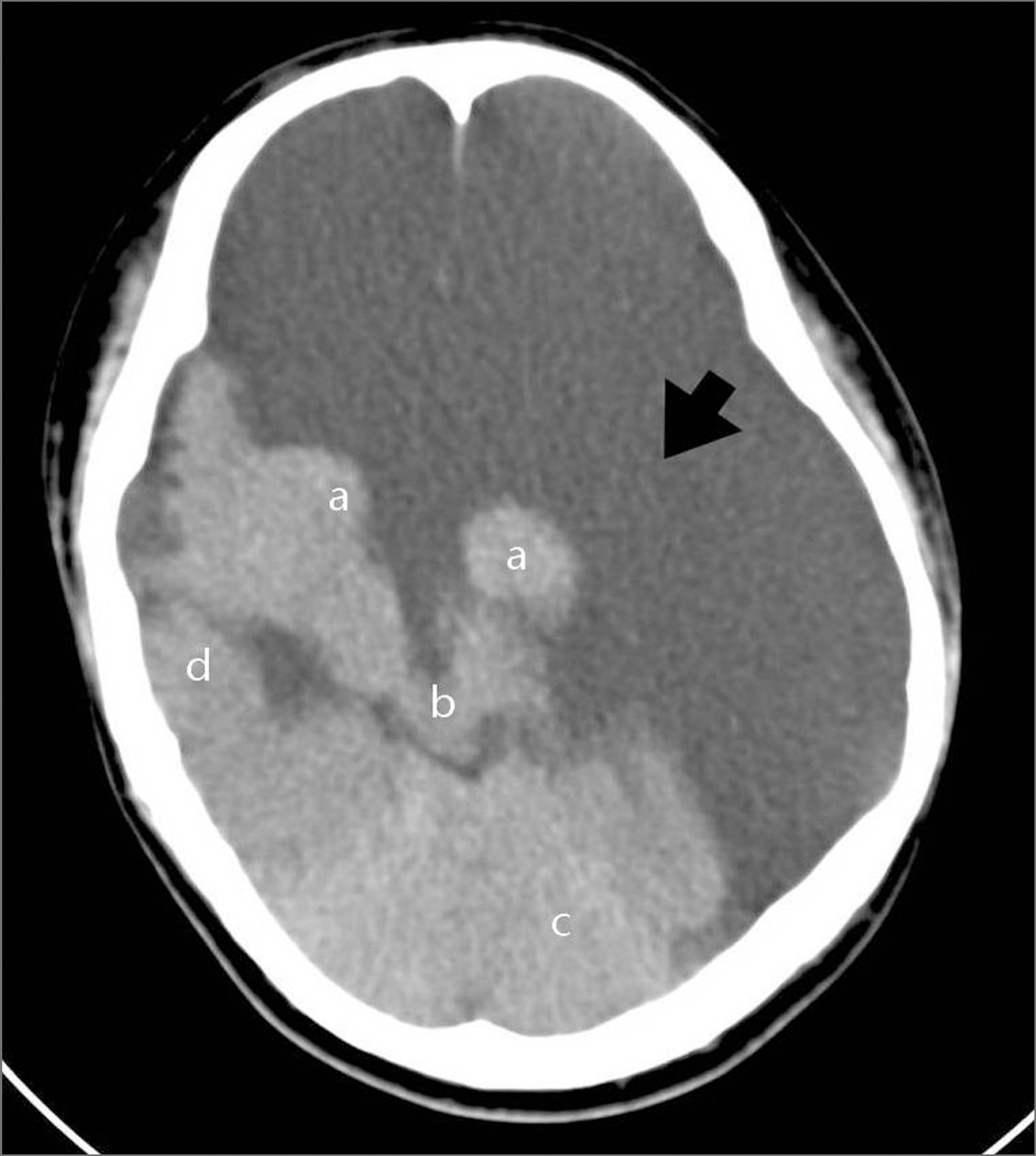
Figure 2. Computed tomogram (CT) of the head. Most of the cerebral cortex is replaced by fluid-filled sac (arrow). Residual parenchyma of the thalamus (a), brainstem (b), cerebellum (c) and partial right fronto-temporal lobe (d)is present posteriorly.

Figure 3. The photograph at the primary gaze shows reduced exotropia and well-everted cilia (taken on the 6th postoperative day).
Reference
-
References
1. Herman DC, Bartley GB, Bullock JD. Ophthalmic findings of hydranencephaly. J Pediatr Ophthalmol Strabismus. 1988; 25:106–11.
Article2. Swaiman K, Ashwal S, Ferriero DM. Pediatric Neurology. 3rd ed.St. Louis: Mosby;1999. p. 273–4.3. Menkes J. Textbook of Child Neurology. 5th ed.Baltimore: Williams & Wilkins;1995. p. 307–8.4. Bae JS, Jang MU, Park SS. Prolonged survival to adulthood of an individual with hydranencephaly. Clin Neurol Neurosurg. 2008; 110:307–9.
Article5. Vinken PJ, Bruyn GW, Klawans HL. Handbook of Clinical Neurology. Amsterdam: Elsevier/North-Holand Biomedical Press;1977. p. 661–80.6. Moisier MA, Lieberman MF, Green WR, et al. Hypoplasia of the optic nerve. Arch Ophthalmol. 1978; 96:1437–42.7. Werth R. Cerebral blindness and plasticity of the visual system in children. A review of visual capacities in patients with occipital lesions, hemispherectomy or hydranencephaly. Restor Neurol Neurosci. 2008; 26:377–89.
