Comparison of Drills Used in Endonasal Dacryocystorhinostomy
- Affiliations
-
- 1Department of Ophthalmology, Hanyang University College of Medicine, Seoul, Korea. lyjot@hanyang.ac.kr
- KMID: 2213376
- DOI: http://doi.org/10.3341/jkos.2010.51.4.479
Abstract
- PURPOSE
To evaluate and compare two types of microdrills that are used in endonasal dacryocystorhinostomy.
METHODS
We retrospectively analyzed medical records and video recordings of 51 patients with 65 affected eyes who underwent endonasal dacryocystorhinostomy for treatment of chronic dacryocystitis or primary acquired nasolacrimal duct obstruction at our hospital between 2005 and 2007. All procedures were performed by the same surgeon. The patients were randomly divided into two groups. For patients in group 1 the surgeon used the Diego powered dissector (Gyrus(R)), while patients in group 2 were treated using the Ossepro (Bein Air(R)) microdrill.
RESULTS
The operation success rate of group 1 was 96.6% and of group 2 was 94.2%. This difference was not statistically significant (p>0.05). The mean operation time was longer in group 1 (45.6 min) than in group 2 (65.8 min). These values, along with the mean drill usage times for each group, were significantly different (p=0.03). The mean revolution per minute(RPMs)or the two groups were also significantly different (p=0.05).
CONCLUSIONS
Our results suggest that microdrills used in endonasal dacryocystorhinostomy should have protective shields to minimize mucosal damage, and employ rapid RPMs to efficiently produce bony openings in the thick anterior processes of the maxilla. The tips of microdrills should also be exposed, to better visualize and acquire good operating fields.
MeSH Terms
Figure
-

Figure 1. (A) Diego powered dissector (Gyrus®)has angulated(18˚) shaft and various tips surrounded by protector. (B) Osseopro (Bien air®) microdrill has more bulky handpiece and exposed tip which provides good surgical view.

Figure 2. Diego powered dissector(Gyrus®) and its utility (A) Endoscopic view of endonasal dacryocystorhinostomy by using Diego powered dissector. Inlet figure was magnification of dissector's tip. (B) Diego powered dissector (Gyrus®) power unit. (C) Various tips can be changed.
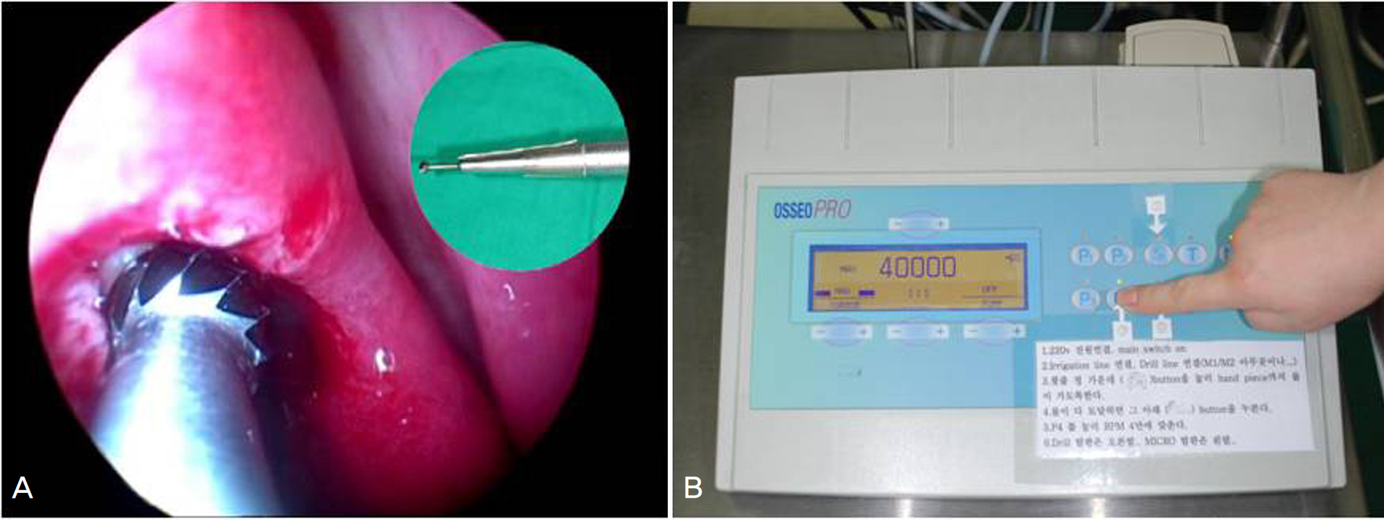
Figure 3. Osseopro (Bien air®) microdrill and its utility (A) Endoscopic view of Osseopro (Bien air®) microdrill in endonasal dacryocystorhinostomy. The tip was fully exposed. Inlet figure shows microdrill's handpiece and tip. (B) Osseopro (Bien air®) microdrill motor unit.

Figure 4. (A) Mechanical injury of inferior nostrils after using Osseopro (Bien air®) (White arrows). (B) Mucosal injury during Osseopro (Bien air®) revolving (Red arrow).
Reference
-
References
1. Yoon SW, Yoon YS, Lee SH. Clinical result of endoscopic abdominal using a microdebrider. Korean J Ophthalmol. 2006; 20:1–6.2. Lee SJ, Na KS, Ji NC. A clinical study on the endonasal microdrill assisted dacryocystorhinostomy. J Korean Ophthalmol Soc. 1998; 39:1620–6.3. Park JD, Kim YI, Shin SG. The factors related to surgical success rate of endonasal dacryocystorhinostomy. J Korean Ophthalmol Soc. 1998; 39:2848–53.4. Kwon YA, Kim HC, Ha MS, et al. Success rates according to the shape of rhinostomy after endonasal dacryocystorhinostomy. J Korean Ophthalmol Soc. 2009; 50:14–8.
Article5. Kwon S, Baek SH. Clinical evaluation of endoscopic endonasal dacryocystorhinostomy. J Korean Ophthalmol Soc. 2004; 45:1403–8.6. Tsirbas A, Wormald PJ. Mechanical endonasal abdominal with mucosal flaps. Br J Ophthalmol. 2003; 87:43–7.7. Lee TS, Kim JS, Cho SH, Choi JS. The surgical result of trans-canalicular LASER-assisted dacryocystothinostomy. J Korean Ophthalmol Soc. 2004; 45:1–7.8. Lester SE, Robson AK, Bearn M. Endoscopic ‘cold steel' versus abdominal dacryocystorhinostomy: completing the audit cycle. J Laryngol Otol. 2008; 122:924–7.9. Tsirbas A, Davis G, Wormald PJ. Mechanical endonasal dacryocystothinostomy versus external dacryocystorhinostomy. Ophthal Plast Reconstr Surg. 2004; 20:50–6.
- Full Text Links
-
- Actions
-
Cited
- CITED
-
- Close
- Share
-
- Similar articles
-
- Success Rate of Endonasal Dacryocystorhinostomy Based on the Location of the Lacrimal Sac
- Endonasal Dacryocystorhinostomy
- Clinical Effecd of Endonasal Lacrimal Surgery
- The Effect of Nasal Cavity Abnormality Related to Surgical Success Rate of Endonasal Dacryocystorhinostomy
- Clinical Consideration of Uncinectomy for Endonasal Dacryocystorhinostomy
