J Korean Rheum Assoc.
2010 Mar;17(1):98-99. 10.4078/jkra.2010.17.1.98.
Calcific Tendinitis of Flexor Carpi Ulnaris Insertion Site
- Affiliations
-
- 1Division of Rheumatology, Department of Internal Medicine, Konkuk University School of Medicine, Seoul, Korea.
- 2Department of Radiology, Korea University College of Medicine, Seoul, Korea.
- 3Division of Rheumatology, Department of Internal Medicine, Korea University College of Medicine, Seoul, Korea. gsong@kumc.or.kr
- KMID: 2202001
- DOI: http://doi.org/10.4078/jkra.2010.17.1.98
Abstract
- No abstract available.
MeSH Terms
Figure
-
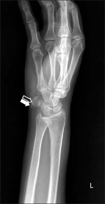
Fig. 1 Oblique view of left wrist plain radiography, showing small irregular bony excrescence on FCU insertion site of the pisifom (black arrow) with calcified nodules around the volar aspect of the pisiform (white arrow).
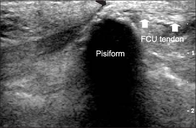
Fig. 2 Left wrist ultrasound showing small irregular calcifications around FCU insertion site of the pisiform (black arrow).
Reference
-
1. Colavita N, Solivetti FM, Vecchioli A, Bock E. Peritendinitis calcarea of flexor carpi ulnaris. Diagn Imaging. 1983. 52:284–286.2. Dilley DF, Tonkin MA. Acute calcific tendinitis in the hand and wrist. J Hand Surg Br. 1991. 16:215–216.3. Moyer RA, Bush DC, Harrington TM. Acute calcific tendinitis of the hand and wrist: a report of 12 cases and a review of the literature. J Rheumatol. 1989. 16:198–202.
- Full Text Links
-
- Actions
-
Cited
- CITED
-
- Close
- Share
-
- Similar articles
-
- Tendon Problems of the Ulnar Wrist
- Successive Acute Calcific Tendinitis at Different Sites
- Surgical Treatment of Chronic Flexor Carpi Ulnaris Tendinopathy
- Surgical Management of Pisiform Bone Deformity Associated with Tendonitis of Flexor Carpi Ulnaris
- Isolated calcific tendinitis at the posterosuperior labrum: a rare case study
