Granular Cell Tumor of the Intradural Extramedullary Spinal Cord : Report of Two Cases with Respect to Radiological Differential Diagnosis
- Affiliations
-
- 1Department of Neurosurgery, Spine Center, Seoul National University Bundang Hospital, Seoul National University College of Medicine, Seongnam, Korea. neurospine@snubh.org
- 2Department of Radiology, Spine Center, Seoul National University Bundang Hospital, Seoul National University College of Medicine, Seongnam, Korea.
- 3Department of Neurosurgery, Asan Medical Center, University of Ulsan College of Medicine, Seoul, Korea.
- KMID: 2190691
- DOI: http://doi.org/10.3340/jkns.2013.53.2.121
Abstract
- Granular cell tumors (GrCTs) of the spinal cord are rare benign tumors with a high rate of local recurrence. Only 6 cases of spinal GrCTs have been reported. GrCT is difficult to distinguish from other benign tumors such as schwannoma using imaging. A radiological "speckled dots" sign may be a useful differentiating feature of GrCT based upon experience with two cases and a review of the literature.
Keyword
MeSH Terms
Figure
-
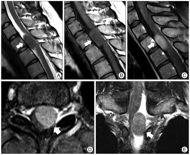
Fig. 1 Preoperative magnetic resonance (MR) images. A : Midsagittal T2-weighted fat-suppressed MR images show an isosignal intensity mass at T1-2. The spinal cord is displaced to the right posterior side. At the center of the tumor, there are low signal speckled dots. B and C : Mid-sagittal T1-weighted MR image (B) and gadolinium-enhanced fat-suppressed MR images (C) reveal isosignal intensity and a homogenous well-enhanced tumor. Speckled dots are observed at the center of the tumor. There is no dura tail sign. D : Axial MR image shows a well-circumscribed tumor displaced to the left side. It shows no extension to the foramen. E : Coronal MR image reveals that the tumor is located eccentrically in the intradural extramedullary and compresses the spinal cord.
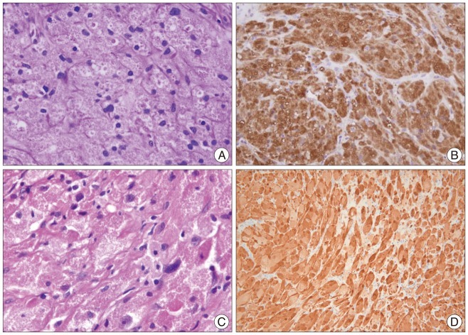
Fig. 2 Histologic examination. A and C : Cases 1 (A) and 2 (C) show granular cells with abundant eosinophilic granules in the cytoplasm. Hyalinizing fibrosis is also observed in all cases. Some lymphocytes have infiltrated the tumor. There is mild nuclear pleomorphism but no necrosis. B and D : The tumor cells are strongly and diffusely immunoreactive with S-100 protein in Case 1 (B) and Case 2 (D).
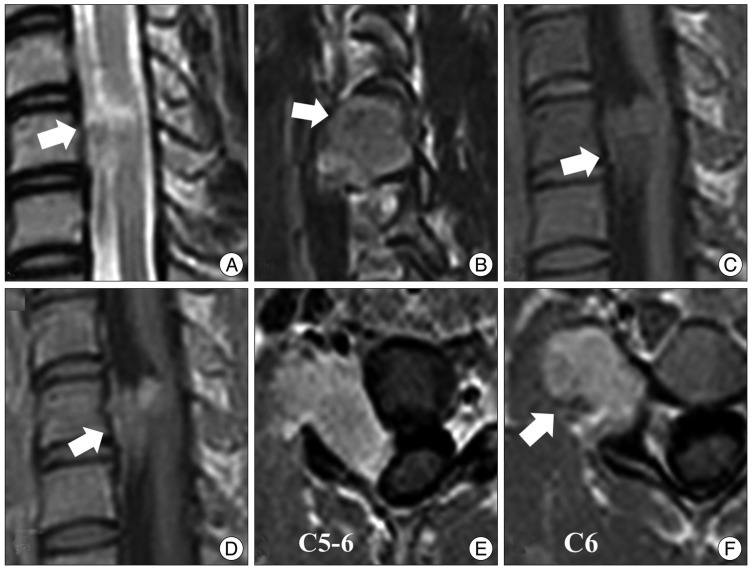
Fig. 3 Preoperative magnetic resonance (MR) images. A : A T2-weighted MR image shows an isosignal intensity mass at C5-6. B : A T2-sagittal MR image shows a tumor extending to the right foramen. Speckled dots in the center of the tumor are of low signal intensity in a T2-weighted MR image (arrow). C and D : T1-weighted MR images show an isointense and well-enhanced mass. E and F : Axial MR images show that the spinal cord is displaced to the left by the tumor. The tumor is mainly located in the extraforaminal area. Low signal speckled dots are observed in all sequences of MR images.
Reference
-
1. Buley ID, Gatter KC, Kelly PM, Heryet A, Millard PR. Granular cell tumours revisited. An immunohistological and ultrastructural study. Histopathology. 1988; 12:263–274. PMID: 2452781.
Article2. Critchley GR, Wallis NT, Cowie RA. Granular cell tumour of the spinal cord : case report. Br J Neurosurg. 1997; 11:452–454. PMID: 9474282.3. Fanburg-Smith JC, Meis-Kindblom JM, Fante R, Kindblom LG. Malignant granular cell tumor of soft tissue : diagnostic criteria and clinicopathologic correlation. Am J Surg Pathol. 1998; 22:779–794. PMID: 9669341.4. Kaiserling E, Ruck P, Xiao JC. Congenital epulis and granular cell tumor : a histologic and immunohistochemical study. Oral Surg Oral Med Oral Pathol Oral Radiol Endod. 1995; 80:687–697. PMID: 8680977.5. Markesbery WR, Duffy PE, Cowen D. Granular cell tumors of the central nervous system. J Neuropathol Exp Neurol. 1973; 32:92–109. PMID: 4346659.
Article6. Ordóñez NG, Mackay B. Granular cell tumor : a review of the pathology and histogenesis. Ultrastruct Pathol. 1999; 23:207–222. PMID: 10503740.7. Pipeleers-Marichal M, Goossens A, De Waele B, KlÖppel G. Granular cell tumour of the appendix in a patient irradiated for a rectal carcinoma. Virchows Arch A Pathol Anat Histopathol. 1990; 417:177–180. PMID: 1695039.
Article8. Qu J, Ma J, Luo L, Ai L, Li S, Dai J. Subdural granular cell tumor in thoracic vertebral canal. Neurol India. 2009; 57:679–681. PMID: 19934580.
Article9. Rhee DJ, Choi YL, Suh YL, Park K. Atypical granular cell tumor of the sellar region. J Korean Neurosurg Soc. 2006; 40:459–462.10. Strömblad LG, Brun A, Cameron R, Cronquist S. Spinal granular cell tumor with subarachnoid hemorrhage : case report. Neurosurgery. 1987; 21:230–233. PMID: 2821449.
- Full Text Links
-
- Actions
-
Cited
- CITED
-
- Close
- Share
-
- Similar articles
-
- Intradural Extramedullary Spinal Ependymoma: A Case Report of Malignant Transformation Occurring
- MR Imaging of Intradural Extramedullary Tuberculoma of the Spinal Cord: Report of Two Cases
- A Case of Intradural, Extramedullary Tuberculous Granuloma Developed During the Treatment of Tuberculous Meningitis
- Removal of Intradural-Extramedullary Spinal Cord Tumors with Unilateral Limited Laminectomy
- Thoracic Intramedullary Schwannoma: 2 Cases Report
