J Korean Orthop Assoc.
2010 Dec;45(6):433-439. 10.4055/jkoa.2010.45.6.433.
Arthroscopic Treatment for Cartilage Lesions of the Talus
- Affiliations
-
- 1Department of Orthopedic Surgery, Pusan Paik Hospital, College of Medicine, Inje University, Busan, Korea. kimjh8142@hanmail.net
- KMID: 2185490
- DOI: http://doi.org/10.4055/jkoa.2010.45.6.433
Abstract
- PURPOSE
To compare clinical results and to evaluate the factors affecting the clinical results after performing arthroscopic chondroplasty, microfracture, and osteochondral autologus transplantation (OAT) due to a chondral defect of the talus.
MATERIALS AND METHODS
This study enrolled 35 patients (36 cases) diagnosed with a chondral defect of the talus and who could be followed over 12 months after arthroscopic chondroplasty, microfracture, or OAT between March 1998 and December 2007. The arthroscopic chondroplasties were carried out in 14 cases (13 patients), the microfractures were carried out in 12 cases (12 patients) and OAT was carried out in 10 cases (10 patients). The lesion staging used Berndt and Harty classification on simple radiographs and Anderson's classification on magnetic resonance images. Clinical results were evaluated and compared by measuring VAS and AOFAS scores at the time of operation, before the operation, and at the time of follow up. Clinical evaluation included location, size, and stage of each lesion as well as the age of individual patient.
RESULTS
There were 13 medial and 23 lateral lesions. The average size of the chondral defects were 1.9 cm2 (range: 1-4 cm2). According to the classification of Berndt and Harty and Anderson, there were 8 stage II, 21 stage III, and 7 stage IV cases. The average follow up period was 15 months (range: 12-30 months). VAS and AOFAS scores showed significant improvement in all treatment groups. However, clinical results according to the operative methods did not show any differences. Lesion size, stage and location, as well as of age of patient had no significant impact on clinical results.
CONCLUSION
We concluded that all three procedures, arthroscopic chondroplasty, microfracture, and OAT, are useful for treating a chondral defect of talus. Location of lesion, size, stage and age of patient did not make a significant difference.
Keyword
MeSH Terms
Figure
-
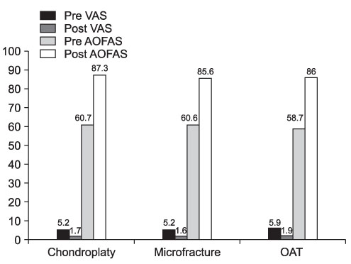
Figure 1 VAS score and AOFAS score showed significant improvement in all treatment groups. However, clinical results according to the operative methods showed no difference. OAT, osteochondral autologous transplantation.
Reference
-
1. Curl WW, Krome J, Gordon ES, Rushing J, Smith BP, Poehling GG. Cartilage injuries: a review of 31,516 knee arthroscopies. Arthroscopy. 1997. 13:456–460.
Article2. Verhagen RA, Struijs PA, Bossuyt PM, van Dijk CN. Systematic review of treatment strategies for osteochondral defects of the talar dome. Foot Ankle Clin. 2003. 8:233–242.
Article3. Al-Shaikh RA, Chou LB, Mann JA, Dreeben SM, Prieskorn D. Autologous osteochondral grafting for talar cartilage defects. Foot Ankle Int. 2002. 23:381–389.
Article4. Assenmacher JA, Kelikian AS, Gottlob C, Kodros S. Arthro-scopically assisted autologous osteochondral transplantation for osteochondral lesions of the talar dome: an MRI and clinical follow-up study. Foot Ankle Int. 2001. 22:544–551.
Article5. Becher C, Thermann H. Results of microfracture in the treatment of articular cartilage defects of the talus. Foot Ankle Int. 2005. 26:583–589.
Article6. Choi CH, Cheon YM. Arthroscopic treatment of osteochondritis dissecans of the talus. J Korean Arthrosc Soc. 2002. 6:161–169.7. Ferkel RD, Zanotti RM, Komenda GA, et al. Arthroscopic treatment of chronic osteochondral lesions of the talus: long-term results. Am J Sports Med. 2008. 36:1750–1762.8. Gautier E, Kolker D, Jakob RP. Treatment of cartilage defects of the talus by autologous osteochondral grafts. J Bone Joint Surg Br. 2002. 84:237–244.
Article9. Giannini S, Vannini F. Operative treatment of osteochondral lesions of the talar dome: current concepts review. Foot Ankle Int. 2004. 25:168–175.
Article10. Gobbi A, Francisco RA, Lubowitz JH, Allegra F, Canata G. Osteochondral lesions of the talus: randomized controlled trial comparing chondroplasty, microfracture, and osteochondral autograft transplantation. Arthroscopy. 2006. 22:1085–1092.
Article11. Kim HK, Moran ME, Salter RB. The potential for regeneration of articular cartilage in defects created by chondral shaving and subchondral abrasion. An experimental investigation in rabbits. J Bone Joint Surg Am. 1991. 73:1301–1315.
Article12. Kim KT, Kim JH, Lee S, Cho GH, Choi DJ. Comparison of arthroscopic debridement and multiple drilling for osteochondritis dissecans of the talus. J Korean Arthrosc Soc. 2005. 9:206–213.13. Kumai T, Takakura Y, Higashiyama I, Tamai S. Arthroscopic drilling for the treatment of osteochondral lesions of the talus. J Bone Joint Surg Am. 1999. 81:1229–1235.
Article14. Lee CH, Chao KH, Huang GS, Wu SS. Osteochondral autografts for osteochondritis dissecans of the talus. Foot Ankle Int. 2003. 24:815–822.
Article15. Loomer R, Fisher C, Lloyd-Smith R, Sisler J, Cooney T. Osteochondral lesions of the talus. Am J Sports Med. 1993. 21:13–19.
Article16. Newman AP. Articular cartilage repair. Am J Sports Med. 1998. 26:309–324.
Article17. Parisien JS. Arthroscopic treatment of osteochondral lesions of the talus. Am J Sports Med. 1986. 14:211–217.
Article18. Parisien JS, Vangsness T. Operative arthroscopy of the ankle. Three years' experience. Clin Orthop Relat Res. 1985. 199:46–53.19. Schuman L, Struijs PA, van Dijk CN. Arthroscopic treatment for osteochondral defects of the talus. Results at follow-up at 2 to 11 years. J Bone Joint Surg Br. 2002. 84:364–368.20. Zengerink M, Szerb I, Hangody L, Dopirak RM, Ferkel RD, van Dijk CN. Current concepts: treatment of osteochondral ankle defects. Foot Ankle Clin. 2006. 11:331–359.
Article21. Berndt AL, Harty M. Transchondral fractures (osteochondritis dissecans) of the talus. J Bone Joint Surg Am. 1959. 41-A:988–1020.
Article22. Anderson IF, Crichton KJ, Grattan-Smith T, Cooper RA, Brazier D. Osteochondral fractures of the dome of the talus. J Bone Joint Surg Am. 1989. 71:1143–1152.
Article23. Dipaola JD, Nelson DW, Colville MR. Characterizing osteochondral lesions by magnetic resonance imaging. Arthroscopy. 1991. 7:101–104.
Article24. Zinman C, Wolfson N, Reis ND. Osteochondritis dissecans of the dome of the talus. Computed tomography scanning in diagnosis and follow-up. J Bone Joint Surg Am. 1988. 70:1017–1019.
Article25. Pritsch M, Horoshovski H, Farine I. Arthroscopic treatment of osteochondral lesions of the talus. J Bone Joint Surg Am. 1986. 68:862–865.
Article
- Full Text Links
-
- Actions
-
Cited
- CITED
-
- Close
- Share
-
- Similar articles
-
- Current Updates in Treatment of Osteochondral Lesions of the Talus
- Comparative Study of the Clinical Results between Arthroscopic Multiple Drilling and Autologous Osteochondral Grafting for Osteochondral Lesions of the Talus
- Redomicrofracture as a Treatment for Osteochondral Lesion of Talus after the Failure of Arthroscopic Microfracture
- Operative Treatment of Osteochondral Lesion of the Talus: Arthroscopic Bone Marrow Stimulation (Multiple Drilling or Microfracture)
- Arthroscopic Treatment for an Osteochondral Lesion of the Talus
