J Cardiovasc Ultrasound.
2014 Mar;22(1):28-31. 10.4250/jcu.2014.22.1.28.
Coronary Artery Fistula Draining into the Left Ventricle
- Affiliations
-
- 1Division of Cardiology, Asan Medical Center, University of Ulsan College of Medicine, Seoul, Korea. jmsong@amc.seoul.kr
- KMID: 2177453
- DOI: http://doi.org/10.4250/jcu.2014.22.1.28
Abstract
- We present a case of 48-year-old male who presented with coronary artery fistula draining into left ventricle. Transthoracic echocardiography showed abnormal blood flow draining into left ventricle, with enlarged coronary arteries and multiple vascular structures around ventricular myocardium. Coronary computed tomography revealed dilatation of entire left coronary artery which was wrapping around left ventricle, and draining into the posterior side of left ventricle. He did not undergo any invasive treatment, because he was not symptomatic.
MeSH Terms
Figure
-
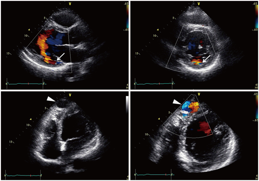
Fig. 1 Abnormal flow draining into the basal posterior portion of left ventricle (arrows) and dilated large echo-free vascular structure (arrowheads), revealed by transthoracic echocardiography and color Doppler images.

Fig. 2 Coronary computed tomography showed markedly dilated and serpentine whole left coronary arteries. LAD: left anterior descending artery, LCX: left circumflex artery.
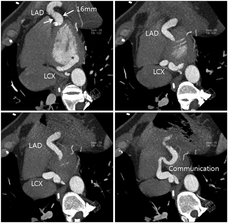
Fig. 3 Coronary computed tomography revealed dilated left coronary arteries and the drainage site to the left ventricle (asterisk). LAD: left anterior descending artery, LCX: left circumflex artery.
Reference
-
1. Ata Y, Turk T, Bicer M, Yalcin M, Ata F, Yavuz S. Coronary arteriovenous fistulas in the adults: natural history and management strategies. J Cardiothorac Surg. 2009; 4:62.
Article2. Fernandes ED, Kadivar H, Hallman GL, Reul GJ, Ott DA, Cooley DA. Congenital malformations of the coronary arteries: the Texas Heart Institute experience. Ann Thorac Surg. 1992; 54:732–740.
Article3. Sommer RJ, Hijazi ZM, Rhodes JF Jr. Pathophysiology of congenital heart disease in the adult: part I: Shunt lesions. Circulation. 2008; 117:1090–1099.
Article4. Mangukia CV. Coronary artery fistula. Ann Thorac Surg. 2012; 93:2084–2092.
Article5. Mohanty SK, Ramanathan KR, Banakal S, Muralidhar K, Kumar P. An interesting case of coronary cameral fistula. Ann Card Anaesth. 2005; 8:152–154.6. Roberts WC. Major anomalies of coronary arterial origin seen in adulthood. Am Heart J. 1986; 111:941–963.
Article7. Vavuranakis M, Bush CA, Boudoulas H. Coronary artery fistulas in adults: incidence, angiographic characteristics, natural history. Cathet Cardiovasc Diagn. 1995; 35:116–120.
Article8. Stierle U, Giannitsis E, Sheikhzadeh A, Potratz J. Myocardial ischemia in generalized coronary artery-left ventricular microfistulae. Int J Cardiol. 1998; 63:47–52.
Article9. Valente AM, Lock JE, Gauvreau K, Rodriguez-Huertas E, Joyce C, Armsby L, Bacha EA, Landzberg MJ. Predictors of long-term adverse outcomes in patients with congenital coronary artery fistulae. Circ Cardiovasc Interv. 2010; 3:134–139.
Article10. Schumacher G, Roithmaier A, Lorenz HP, Meisner H, Sauer U, Müller KD, Sebening F, Bühlmeyer K. Congenital coronary artery fistula in infancy and childhood: diagnostic and therapeutic aspects. Thorac Cardiovasc Surg. 1997; 45:287–294.
Article
- Full Text Links
-
- Actions
-
Cited
- CITED
-
- Close
- Share
-
- Similar articles
-
- A case report of coronary artery fistula to the left ventricle
- Surgical Management of a Large Right Coronary Artery to Left Ventricle Fistula in a Neonate: A Case Report
- Three Cases of Coronary Artery Fistula from Right Coronay to Left Ventricle
- Five Cases of Coronary Arteriovenous Fistula from Coronary Artery to Left Ventricle
- Congenital Giant Left Circumflex Artery-to-Left Ventricle Fistula Detected Using Two-Dimensional and Doppler Echocardiography
