Chonnam Med J.
2015 Aug;51(2):91-96. 10.4068/cmj.2015.51.2.91.
Factors Affecting the Accuracy of Intraocular Lens Power Calculation with Lenstar
- Affiliations
-
- 1Department of Ophthalmology, Chonnam National University Medical School and Hospital, Gwangju, Korea. kcyoon@jnu.ac.kr
- KMID: 2172137
- DOI: http://doi.org/10.4068/cmj.2015.51.2.91
Abstract
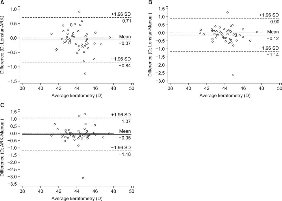
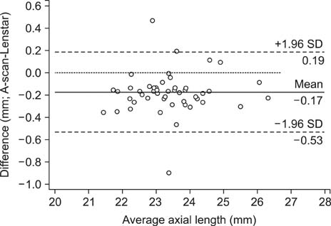
- This retrospective study was performed to compare refractive outcomes measured by conventional methods and by use of the Lenstar biometer and to investigate the factors affecting intraocular lens (IOL) power calculation with Lenstar with and without IOL-constant optimization. The study included 100 eyes of 86 patients who underwent cataract surgery. Corneal curvature was measured with a manual keratometer (MK), automated keratometer (AK), and the Lenstar biometer, and axial length (AL) was measured by A-scan and Lenstar. Mean numerical error (MNE) and mean absolute error (MAE) were compared between AK and MK with A-scan, and Lenstar with and without optimization. Factors affecting the accuracy of the IOL power calculation by use of Lenstar with and without optimization were analyzed. No significant differences were observed in the MNE or MAE among the devices. The proportion of MAE within 0.5 D was higher for Lenstar with optimization (62.7%) than without optimization (46.2%). The proportion of MAE within 0.5 D was 62% and 58% for MK and AK with A-scan, respectively. Without optimization, the MAE was smaller in eyes with ALs between 23 mm and 25 mm (p=0.03), whereas it was smaller at higher corneal powers when the IOL constant was optimized (>44 D, p=0.03). The IOL power calculations showed no significant differences among the devices, but the results of MAE within 0.5 D by use of Lenstar without optimization were worse than those of conventional methods. The AL influenced the accuracy of refractive outcomes determined by using Lenstar without optimization, and corneal curvature was shown to affect the accuracy of refractive measurements using Lenstar with optimization.
MeSH Terms
Figure
Reference
-
1. Connors R 3rd, Boseman P 3rd, Olson RJ. Accuracy and reproducibility of biometry using partial coherence interferometry. J Cataract Refract Surg. 2002; 28:235–238.
Article2. Rajan MS, Keilhorn I, Bell JA. Partial coherence laser interferometry vs conventional ultrasound biometry in intraocular lens power calculations. Eye (Lond). 2002; 16:552–556.
Article3. Song BY, Yang KJ, Yoon KC. Accuracy of partial coherence interferometry in intraocular lens power calculation. J Korean Ophthalmol Soc. 2005; 46:775–780.4. Findl O, Drexler W, Menapace R, Heinzl H, Hitzenberger CK, Fercher AF. Improved prediction of intraocular lens power using partial coherence interferometry. J Cataract Refract Surg. 2001; 27:861–867.
Article5. Eleftheriadis H. IOLMaster biometry: refractive results of 100 consecutive cases. Br J Ophthalmol. 2003; 87:960–963.
Article6. Haigis W, Lege B, Miller N, Schneider B. Comparison of immersion ultrasound biometry and partial coherence interferometry for intraocular lens calculation according to Haigis. Graefes Arch Clin Exp Ophthalmol. 2000; 238:765–773.
Article7. Kiss B, Findl O, Menapace R, Wirtitsch M, Petternel V, Drexler W, et al. Refractive outcome of cataract surgery using partial coherence interferometry and ultrasound biometry: clinical feasibility study of a commercial prototype II. J Cataract Refract Surg. 2002; 28:230–234.
Article8. Buckhurst PJ, Wolffsohn JS, Shah S, Naroo SA, Davies LN, Berrow EJ. A new optical low coherence reflectometry device for ocular biometry in cataract patients. Br J Ophthalmol. 2009; 93:949–953.
Article9. Jasvinder S, Khang TF, Sarinder KK, Loo VP, Subrayan V. Agreement analysis of LENSTAR with other techniques of biometry. Eye (Lond). 2011; 25:717–724.
Article10. Madge SN, Khong CH, Lamont M, Bansal A, Antcliff RJ. Optimization of biometry for intraocular lens implantation using the Zeiss IOLMaster. Acta Ophthalmol Scand. 2005; 83:436–438.
Article11. Nemeth G, Nagy A, Berta A, Modis L Jr. Comparison of intraocular lens power prediction using immersion ultrasound and optical biometry with and without formula optimization. Graefes Arch Clin Exp Ophthalmol. 2012; 250:1321–1325.
Article12. Aristodemou P, Knox Cartwright NE, Sparrow JM, Johnston RL. Intraocular lens formula constant optimization and partial coherence interferometry biometry: Refractive outcomes in 8108 eyes after cataract surgery. J Cataract Refract Surg. 2011; 37:50–62.
Article13. Hsieh YT, Wang IJ. Intraocular lens power measured by partial coherence interferometry. Optom Vis Sci. 2012; 89:1697–1701.
Article14. Jo DH, Oh JY, Kim MK, Lee JH, Wee WR. Corneal power estimation using orbscan II videokeratography in eyes with previous corneal refractive surgeries. J Korean Ophthalmol Soc. 2009; 50:1730–1734.
Article15. Tappeiner C, Rohrer K, Frueh BE, Waelti R, Goldblum D. Clinical comparison of biometry using the non-contact optical low coherence reflectometer (Lenstar LS 900) and contact ultrasound biometer (Tomey AL-3000) in cataract eyes. Br J Ophthalmol. 2010; 94:666–667.
Article16. Olsen T. Prediction of the effective postoperative (intraocular lens) anterior chamber depth. J Cataract Refract Surg. 2006; 32:419–424.
Article17. Gale RP, Saldana M, Johnston RL, Zuberbuhler B, McKibbin M. Benchmark standards for refractive outcomes after NHS cataract surgery. Eye (Lond). 2009; 23:149–152.
Article18. Aristodemou P, Knox Cartwright NE, Sparrow JM, Johnston RL. Formula choice: Hoffer Q, Holladay 1, or SRK/T and refractive outcomes in 8108 eyes after cataract surgery with biometry by partial coherence interferometry. J Cataract Refract Surg. 2011; 37:63–71.
Article19. Ueda T, Ikeda H, Ota T, Matsuura T, Hara Y. Relationship between postoperative refractive outcomes and cataract density: multiple regression analysis. J Cataract Refract Surg. 2010; 36:806–809.
Article20. Prinz A, Neumayer T, Buehl W, Kiss B, Sacu S, Drexler W, et al. Influence of severity of nuclear cataract on optical biometry. J Cataract Refract Surg. 2006; 32:1161–1165.
Article21. Häsemeyer S, Hugger P, Jonas JB. Preoperative biometry of cataractous eyes using partial coherence laser interferometry. Graefes Arch Clin Exp Ophthalmol. 2003; 241:251–252.
Article22. Kim SM, Choi J, Choi S. Refractive predictability of partial coherence interferometry and factors that can affect it. Korean J Ophthalmol. 2009; 23:6–12.
Article
- Full Text Links
- Actions
-
Cited
- CITED
-
- Close
- Share
- Similar articles
-
- Intraocular Lens Power Calculation in Cataract Surgery after Excimer Laser Photorefractive Keratectomy
- Accuracy of Partial Coherence Interferometry in Intraocular Lens Power Calculation
- Evaluation for the Accuracy of the SRK/T Formula in PCL Implanted Patients(I)
- Comparison of the SRK and SRKII Formulas with Revision of Constant A in Intraocular Lens Power Calculation
- A Clinical Study of Intraocular Lens Power Calculation


