Cancer Res Treat.
2013 Sep;45(3):239-243.
Angiomatoid Fibrous Histiocytoma as a Second Tumor in a Young Adult with Testicular Cancer
- Affiliations
-
- 1Department of Internal Medicine, Inje University Ilsan Paik Hospital, Inje University College of Medicine, Goyang, Korea. Dr-Yi@paik.ac.kr
- 2Department of Pathology, Inje University Ilsan Paik Hospital, Inje University College of Medicine, Goyang, Korea.
- 3Department of Radiology, Kangbuk Samsung Hospital, Sungkyunkwan University School of Medicine, Seoul, Korea.
Abstract
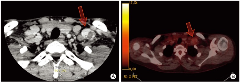
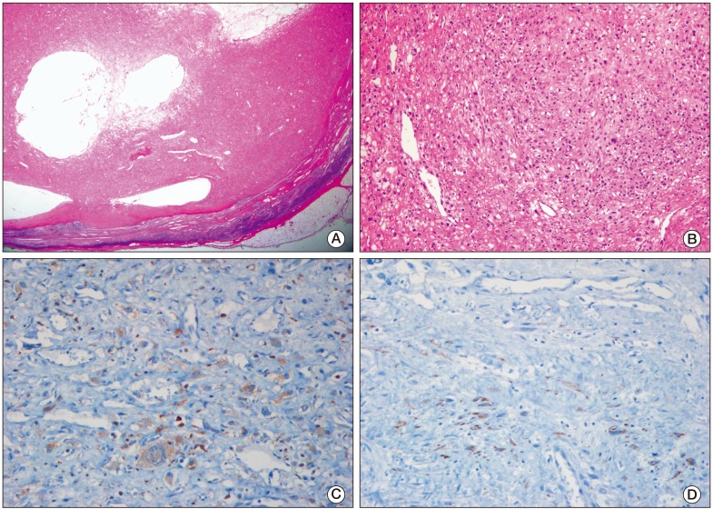
- Angiomatoid fibrous histiocytoma (AFH) is a rare soft tissue tumor, with a low-grade malignant potential, occurring predominantly in children and young adults. Association between AFH and other malignancies has been rarely reported. A 27-year-old man who presented with a palpable abdominal mass was diagnosed as having testicular cancer with multiple liver and lung metastases. At 16 months after chemotherapy, a follow-up computed tomographic scan revealed a supraclavicular mass measuring 3 cm in size, which was suspected to be a recurrence. The patient underwent surgical excision, and the mass was pathologically diagnosed as a AFH. The patient has had no local recurrence and no distant metastasis for 12 months after resection. To the best of our knowledge, this is the first case report of AFH as a second tumor in a patient with testicular cancer.
Keyword
MeSH Terms
Figure
Reference
-
1. Enzinger FM. Angiomatoid malignant fibrous histiocytoma: a distinct fibrohistiocytic tumor of children and young adults simulating a vascular neoplasm. Cancer. 1979; 44:2147–2157. PMID: 228836.2. Costa MJ, Weiss SW. Angiomatoid malignant fibrous histiocytoma: a follow-up study of 108 cases with evaluation of possible histologic predictors of outcome. Am J Surg Pathol. 1990; 14:1126–1132. PMID: 2174650.3. Waters BL, Panagopoulos I, Allen EF. Genetic characterization of angiomatoid fibrous histiocytoma identifies fusion of the FUS and ATF-1 genes induced by a chromosomal translocation involving bands 12q13 and 16p11. Cancer Genet Cytogenet. 2000; 121:109–116. PMID: 11063792.
Article4. Hallor KH, Mertens F, Jin Y, Meis-Kindblom JM, Kindblom LG, Behrendtz M, et al. Fusion of the EWSR1 and ATF1 genes without expression of the MITF-M transcript in angiomatoid fibrous histiocytoma. Genes Chromosomes Cancer. 2005; 44:97–102. PMID: 15884099.5. Rossi S, Szuhai K, Ijszenga M, Tanke HJ, Zanatta L, Sciot R, et al. EWSR1-CREB1 and EWSR1-ATF1 fusion genes in angiomatoid fibrous histiocytoma. Clin Cancer Res. 2007; 13:7322–7328. PMID: 18094413.
Article6. Pettinato G, Manivel JC, De Rosa G, Petrella G, Jaszcz W. Angiomatoid malignant fibrous histiocytoma: cytologic, immunohistochemical, ultrastructural, and flow cytometric study of 20 cases. Mod Pathol. 1990; 3:479–487. PMID: 2170972.7. Gambini C, Haupt R, Rongioletti F. Angiomatoid (malignant) fibrous histiocytoma as a second tumour in a child with neuroblastoma. Br J Dermatol. 2000; 142:537–539. PMID: 10735967.
Article8. Fanburg-Smith JC, Miettinen M. Angiomatoid "malignant" fibrous histiocytoma: a clinicopathologic study of 158 cases and further exploration of the myoid phenotype. Hum Pathol. 1999; 30:1336–1343. PMID: 10571514.
Article9. Martelli L, Collini P, Meazza C, Bianchi A, Salvini F, Zaffignani E, et al. Angiomatoid fibrous histiocytoma in an HIV-positive child. J Pediatr Hematol Oncol. 2008; 30:242–244. PMID: 18376290.
Article10. Coleman MP, Gatta G, Verdecchia A, Esteve J, Sant M, Storm H, et al. EUROCARE-3 summary: cancer survival in Europe at the end of the 20th century. Ann Oncol. 2003; 14(Suppl 5):v128–v149. PMID: 14684503.
Article11. Becherer A, De Santis M, Karanikas G, Szabo M, Bokemeyer C, Dohmen BM, et al. FDG PET is superior to CT in the prediction of viable tumour in post-chemotherapy seminoma residuals. Eur J Radiol. 2005; 54:284–288. PMID: 15837411.
Article12. Oechsle K, Hartmann M, Brenner W, Venz S, Weissbach L, Franzius C, et al. [18F]Fluorodeoxyglucose positron emission tomography in nonseminomatous germ cell tumors after chemotherapy: the German multicenter positron emission tomography study group. J Clin Oncol. 2008; 26:5930–5935. PMID: 19018083.
Article13. Travis LB, Fossa SD, Schonfeld SJ, McMaster ML, Lynch CF, Storm H, et al. Second cancers among 40,576 testicular cancer patients: focus on long-term survivors. J Natl Cancer Inst. 2005; 97:1354–1365. PMID: 16174857.
Article14. Grossman LD, White RR 4th, Arber DA. Angiomatoid fibrous histiocytoma. Ann Plast Surg. 1996; 36:649–651. PMID: 8792977.
Article15. Costa MA, Silva I, Carvalhido L, Azevedo I, Alves L, Leal C, et al. Angiomatoid fibrous histiocytoma of the arm treated by radiotherapy for local recurrence: case report. Med Pediatr Oncol. 1997; 28:373–376. PMID: 9121405.


