Three-dimensional imaging of progressive facial hemiatrophy (Parry-Romberg syndrome) with unusual conjunctival findings
- Affiliations
-
- 1Department of Oral Medicine and Radiology, AB Shetty Memorial Institute of Dental Sciences, Nitte University, Mangalore, India. p_preethidr@rediffmail.com
- KMID: 2167405
- DOI: http://doi.org/10.5624/isd.2011.41.4.183
Abstract
- Progressive hemifacial atrophy, also known as Parry-Romberg syndrome, is an uncommon degenerative condition which is poorly defined. It is characterized by a slow and progressive atrophy affecting one side of the face. The onset usually occurs during the first two decades of life. Characteristically, the atrophy progresses slowly for several years, and then it becomes stable. Ophthalmic involvement is common, with progressive enophthalmos which is a frequent finding. Cutaneous pigmentation is common in such conditions, however its extension to the conjunctiva is rarely reported. We report a case of Parry Romberg syndrome with characteristic clinical and radiographic presentation accompanied with rare ocular findings. The clinical features, radiological findings, and differential diagnoses to be considered, and the available treatment options are discussed in this report.
MeSH Terms
Figure
-
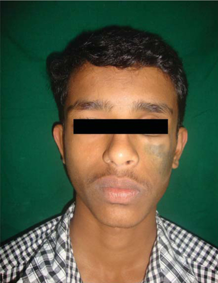
Fig. 1 Clinical photograph of the patient shows marked hypoplasia of the left malar area with diffuse blackish pigmentation of the same region.
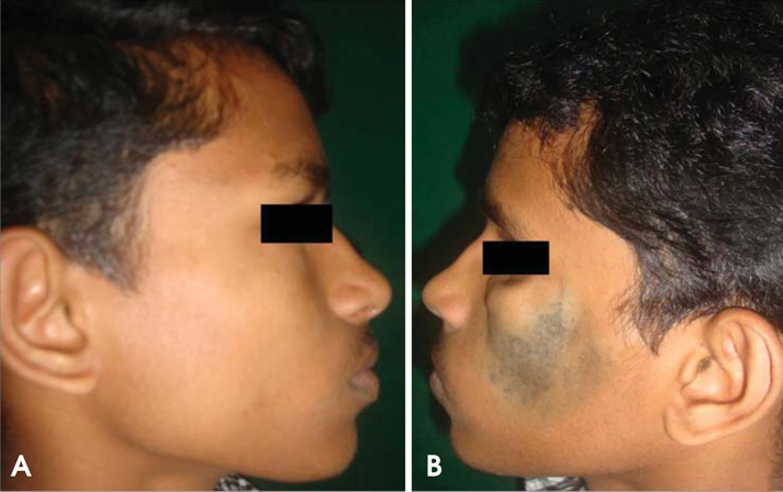
Fig. 2 Photographs compare the normal and abnormal sides.
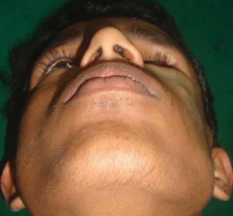
Fig. 3 Photograph shows enophthalmos and flattening of zygomatic complex.
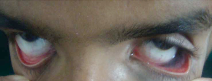
Fig. 4 Photograph shows the pigmentation of the left palpebral conjunctiva.
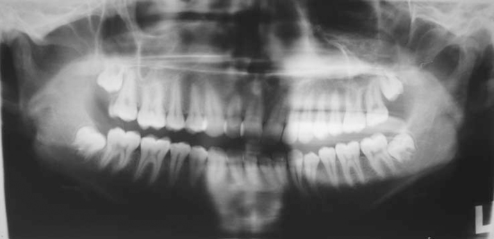
Fig. 5 Panoramic radiograph shows shorter crowns and roots of teeth on the left side compared with the right side.
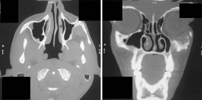
Fig. 6 Axial and coronal CT images reveal the hypoplastic left zygomatic complex and maxillary sinus.
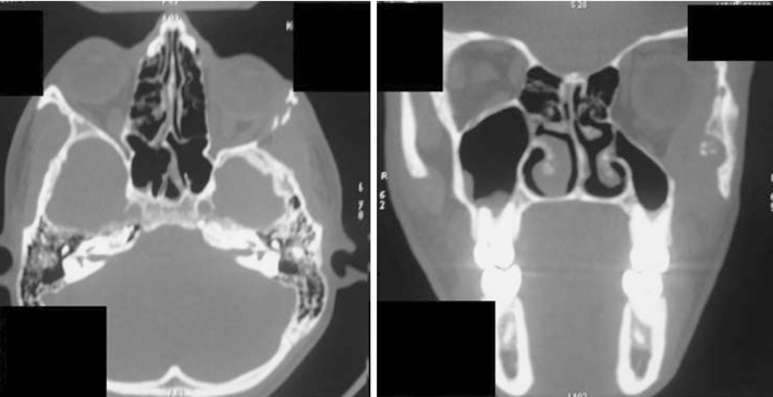
Fig. 7 Axial CT image shows the left eye ball at a lower level when compared to right side. Also observe the hypoplastic lateral orbital wall. Also coronal CT image reveals the shrunken left eyeball, hypoplastic maxillary sinus and lateral orbital wall.
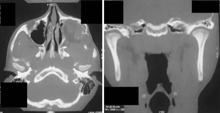
Fig. 8 Coronal and axial CT images show the minimal condylar changes on the right side compared with the left side.
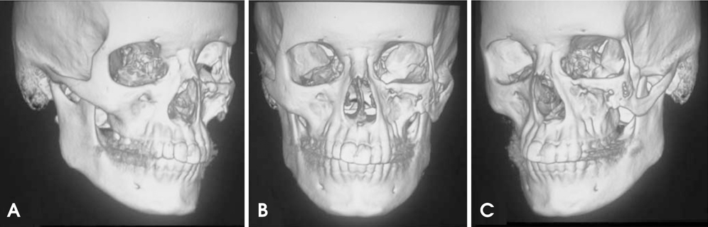
Fig. 9 3D CT images show the normal anatomical features on the right side (A), deviated nasal septum and tilt in the occlusal table in the frontal view (B), hypoplastic zygomatic complex and orbital wall on left side (C).
Reference
-
1. Stone J. Parry-Romberg syndrome. Pract Neurol. 2006. 6:185–188.
Article2. Rangare AL, Babu SG, Thomas PS, Shetty SR. Parry-Romberg syndrome: a rare case report. J Oral Maxillofac Res. 2011. 2:e5.
Article3. Pinheiro TP, Silva CC, Silveira CS, Botelho PC, Pinheiro MG, Pinheiro Jde J. Progressive hemifacial atrophy - case report. Med Oral Patol Oral Cir Bucal. 2006. 11:E112–E114.4. Hakin KN, Yokoyama C, Wright JE. Hemifacial atrophy: an unusual cause of enophthalmos. Br J Ophthalmol. 1990. 74:496–497.
Article5. Kumar AA, Kumar RA, Shantha GP, Aloogopinathan G. Progressive hemi facial atrophy - Parry Romberg syndrome presenting as severe facial pain in a young man: a case report. Cases J. 2009. 2:6776.
Article6. Miller MT, Sloane H, Goldberg MF, Grisolano J, Frenkel M, Mafee MF. Progressive hemifacial atrophy (Parry-Romberg disease). J Pediatr Ophthalmol Strabismus. 1987. 24:27–36.
Article7. Mazzeo N, Fisher JG, Mayer MH, Mathieu GP. Progressive hemifacial atrophy (Parry-Romberg syndrome). Case report. Oral Surg Oral Med Oral Pathol Oral Radiol Endod. 1995. 79:30–35.
Article8. Cory RC, Clayman DA, Faillace WJ, McKee SW, Gama CH. Clinical and radiologic findings in progressive facial hemiatrophy (Parry-Romberg syndrome). AJNR Am J Neuroradiol. 1997. 18:751–757.9. Chbicheb M, Gelot A, Rivier F, Roubertie A, Humbertclaude V, Coubes P, et al. Parry-Romberg's syndrome and epilepsy. Rev Neurol. 2005. 161:92–97.10. de la Fuente A, Jimenez A. Latissimus dorsi free flap for restoration of facial contour defects. Ann Plast Surg. 1989. 22:1–8.
Article11. Donofrio LM. Panfacial volume restoration with fat. Dermatol Surg. 2005. 31:1496–1505.
Article
