J Korean Med Sci.
2015 Apr;30(4):475-482. 10.3346/jkms.2015.30.4.475.
Baseline Predictors of Visual Acuity and Retinal Thickness in Patients with Retinal Vein Occlusion
- Affiliations
-
- 1Department of Ophthalmology, Samsung Medical Center, Sungkyunkwan University School of Medicine, Seoul, Korea.
- 2Department of Ophthalmology, Asan Medical Center, University of Ulsan, Seoul, Korea. yhyoon@amc.seoul.kr
- 3Department of Ophthalmology, Kangnam Sacred Heart Hospital, Hallym University, Seoul, Korea.
- 4Department of Ophthalmology, St. Mary's Eye Clinic, Busan, Korea.
- 5Department of Ophthalmology, Seoul National University Bundang Hospital, Seongnam, Korea.
- 6Department of Ophthalmology, Chungnam National University Hospital, Deajeon, Korea.
- KMID: 2164457
- DOI: http://doi.org/10.3346/jkms.2015.30.4.475
Abstract
- This study investigated the baseline predictors of best corrected visual acuity (BCVA) and central retinal thickness (CRT) at 6 months in patients with treatment-naive branch retinal vein occlusion (BRVO) and central retinal vein occlusion (CRVO). This multicenter, interventional case series included 208 BRVO and 123 CRVO patients with follow-up period of 6 months or more. Outcome measures of BCVA (logMAR) included absolute change from baseline and a gain or loss of > or = 0.3 from baseline. Outcome measures of CRT included absolute change from baseline and a measurement of < or = 250 microm or > or = 400 microm at 6 months. Univariate and multiple regression analyses were done to find baseline predictors. For BRVO, younger age, worse baseline BCVA, and shorter duration of symptom were associated with more gain in BCVA. For CRVO, worse baseline BCVA was associated with more gain in BCVA. For CRT outcomes, higher baseline CRT predicted greater decrease at 6 months in both BRVO and CRVO. Younger age and better baseline BCVA were associated with an increased likelihood of measurement of a < or = 250 microm outcome for BRVO and CRVO, respectively. For CRVO, smoking was associated with greater decrease from baseline and decreased likelihood of measurement of a CRT > or = 400 microm at 6 months. In conclusion, several baseline factors including age, symptom duration, and baseline BCVA and CRT are associated with BCVA and CRT outcomes at 6 months, which may help to predict disease course for RVO patients.
Keyword
MeSH Terms
Figure
-
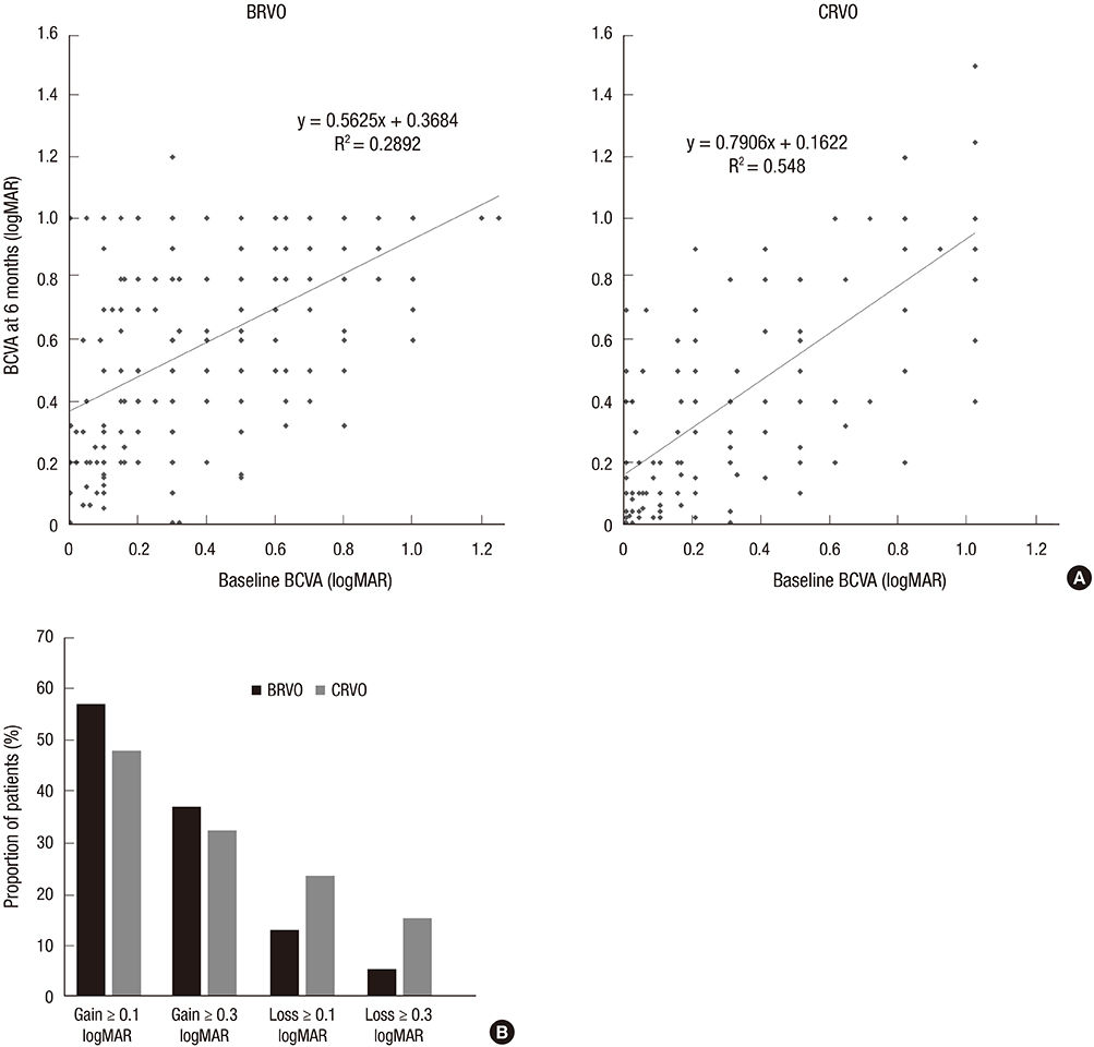
Fig. 1 Best-corrected visual acuity (BCVA) of patients with branch retinal vein occlusion (BRVO) and central retinal vein occlusion (CRVO). (A) Distribution of BCVA (decimal) at baseline and 6 months in patients with BRVO and CRVO. (B) Summary of changes in BCVA from baseline in patients with BRVO and CRVO.
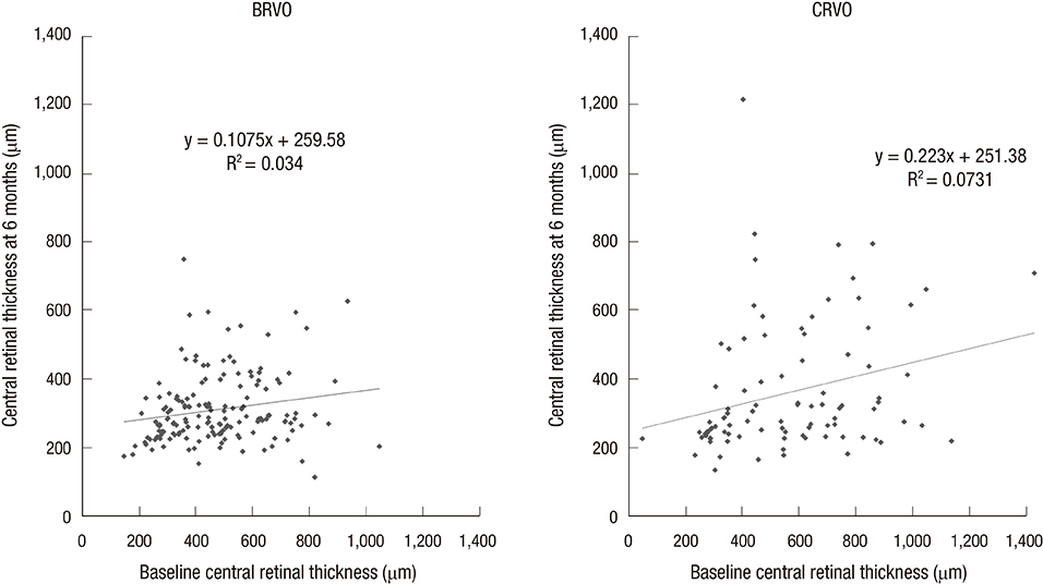
Fig. 2 Distribution of central retinal thickness at baseline and 6 months in patients with branch retinal vein occlusion (BRVO) and central retinal vein occlusion (CRVO).
Cited by 1 articles
-
Significance of Early Visual Responses to Bevacizumab for Macular Edema in Branch Retinal Vein Occlusion
Gahyung Ryu, Donghyoun Noh, Junyeop Lee, Min Sagong
J Korean Ophthalmol Soc. 2017;58(8):937-946. doi: 10.3341/jkos.2017.58.8.937.
Reference
-
1. Wong TY, Scott IU. Clinical practice. Retinal-vein occlusion. N Engl J Med. 2010; 363:2135–2144.2. Rogers SL, McIntosh RL, Lim L, Mitchell P, Cheung N, Kowalski JW, Nguyen HP, Wang JJ, Wong TY. Natural history of branch retinal vein occlusion: an evidence-based systematic review. Ophthalmology. 2010; 117:1094–1101.3. McIntosh RL, Rogers SL, Lim L, Cheung N, Wang JJ, Mitchell P, Kowalski JW, Nguyen HP, Wong TY. Natural history of central retinal vein occlusion: an evidence-based systematic review. Ophthalmology. 2010; 117:1113–1123.e15.4. Campochiaro PA, Heier JS, Feiner L, Gray S, Saroj N, Rundle AC, Murahashi WY, Rubio RG. BRAVO Investigators. Ranibizumab for macular edema following branch retinal vein occlusion: six-month primary end point results of a phase III study. Ophthalmology. 2010; 117:1102–1112.e1.5. Brown DM, Campochiaro PA, Singh RP, Li Z, Gray S, Saroj N, Rundle AC, Rubio RG, Murahashi WY. CRUISE Investigator. Ranibizumab for macular edema following central retinal vein occlusion: six-month primary end point results of a phase III study. Ophthalmology. 2010; 117:1124–1133.e1.6. Haller JA, Bandello F, Belfort R Jr, Blumenkranz MS, Gillies M, Heier J, Loewenstein A, Yoon YH, Jiao J, Li XY, et al. Ozurdex GENEVA Study Group. Dexamethasone intravitreal implant in patients with macular edema related to branch or central retinal vein occlusion twelve-month study results. Ophthalmology. 2011; 118:2453–2460.7. Scott IU, Ip MS, VanVeldhuisen PC, Oden NL, Blodi BA, Fisher M, Chan CK, Gonzalez VH, Singerman LJ, Tolentino M, et al. SCORE Study Research Group. A randomized trial comparing the efficacy and safety of intravitreal triamcinolone with standard care to treat vision loss associated with macular Edema secondary to branch retinal vein occlusion: the Standard Care vs Corticosteroid for Retinal Vein Occlusion (SCORE) study report 6. Arch Ophthalmol. 2009; 127:1115–1128.8. Ip MS, Scott IU, VanVeldhuisen PC, Oden NL, Blodi BA, Fisher M, Singerman LJ, Tolentino M, Chan CK, Gonzalez VH, et al. SCORE Study Research Group. A randomized trial comparing the efficacy and safety of intravitreal triamcinolone with observation to treat vision loss associated with macular edema secondary to central retinal vein occlusion: the Standard Care vs Corticosteroid for Retinal Vein Occlusion (SCORE) study report 5. Arch Ophthalmol. 2009; 127:1101–1114.9. Scott IU, VanVeldhuisen PC, Oden NL, Ip MS, Blodi BA, Hartnett ME, Cohen G. Standard Care versus COrticosteroid for REtinal Vein Occlusion Study Investigator Group. Baseline predictors of visual acuity and retinal thickness outcomes in patients with retinal vein occlusion: Standard Care Versus COrticosteroid for REtinal Vein Occlusion Study report 10. Ophthalmology. 2011; 118:345–352.10. Lee JY, Yoon YH, Kim HK, Yoon HS, Kang SW, Kim JG, Park KH, Jo YJ. Korean RVO Study. Baseline characteristics and risk factors of retinal vein occlusion: a study by the Korean RVO Study Group. J Korean Med Sci. 2013; 28:136–144.11. Kiernan DF, Hariprasad SM. Normative databases in SD-OCT: a status report. accessed on 1 March 2012. Available at http://www.retinalphysician.com/articleviewer.aspx?articleID=104438.12. Heussen FM, Ouyang Y, McDonnell EC, Narala R, Ruiz-Garcia H, Walsh AC, Sadda SR. Comparison of manually corrected retinal thickness measurements from multiple spectral-domain optical coherence tomography instruments. Br J Ophthalmol. 2012; 96:380–385.13. The Branch Vein Occlusion Study Group. Argon laser photocoagulation for macular edema in branch vein occlusion. Am J Ophthalmol. 1984; 98:271–282.14. Ip MS, Oden NL, Scott IU, VanVeldhuisen PC, Blodi BA, Figueroa M, Antoszyk A, Elman M. SCORE Study Investigator Group. SCORE Study report 3: study design and baseline characteristics. Ophthalmology. 2009; 116:1770–1777.e1.15. Klein R, Klein BE, Moss SE, Meuer SM. The epidemiology of retinal vein occlusion: the Beaver Dam Eye Study. Trans Am Ophthalmol Soc. 2000; 98:133–141. discussion 41-3.16. Klein R, Knudtson MD, Lee KE, Gangnon R, Klein BE. The Wisconsin Epidemiologic Study of Diabetic Retinopathy XXIII: the twenty-five-year incidence of macular edema in persons with type 1 diabetes. Ophthalmology. 2009; 116:497–503.17. Thorne JE, Daniel E, Jabs DA, Kedhar SR, Peters GB, Dunn JP. Smoking as a risk factor for cystoid macular edema complicating intermediate uveitis. Am J Ophthalmol. 2008; 145:841–846.
- Full Text Links
-
- Actions
-
Cited
- CITED
-
- Close
- Share
-
- Similar articles
-
- Clinical Features According to the Occlusion Site in Patients with Branch Retinal Vein Occlusion
- Outcome of Vitrectomy on Vitreoretinal Pathologies due to Branch Retinal Vein Occlusion
- A Case of Cilioretinal Artery Occlusion Associated with Central Retinal Vein Occlusion
- The Efficacy of Intravitreal Bevacizumab in the Treatment of Macular Edema
- Clinical Aspect of Branch Retinal Vein Occlusion
