J Korean Neurosurg Soc.
2015 Dec;58(6):539-546. 10.3340/jkns.2015.58.6.539.
Learning Curve of Percutaneous Endoscopic Lumbar Discectomy Based on the Period (Early vs. Late) and Technique (in-and-out vs. in-and-out-and-in): A Retrospective Comparative Study
- Affiliations
-
- 1Department of Neurosurgery, Spine and Spinal Cord Institute, Gangnam Severance Spine Hospital, Yonsei University College of Medicine, Seoul, Korea. ahnsangsoak@hanmail.net
- 2Department of Radiology, Dong-A University Medical Center, Busan, Korea.
- KMID: 2151156
- DOI: http://doi.org/10.3340/jkns.2015.58.6.539
Abstract
OBJECTIVE
To report the learning curve of percutaneous endoscopic lumbar discectomy (PELD) for a surgeon who had not been previously exposed to this procedure based on the period and detailed technique with a retrospective matched comparative design.
METHODS
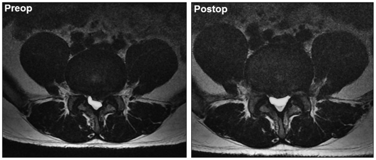
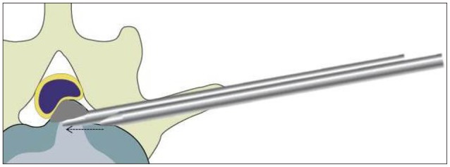
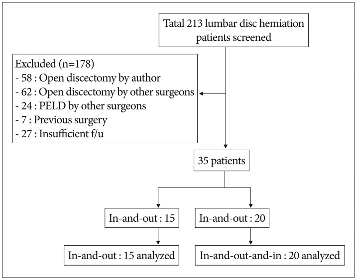
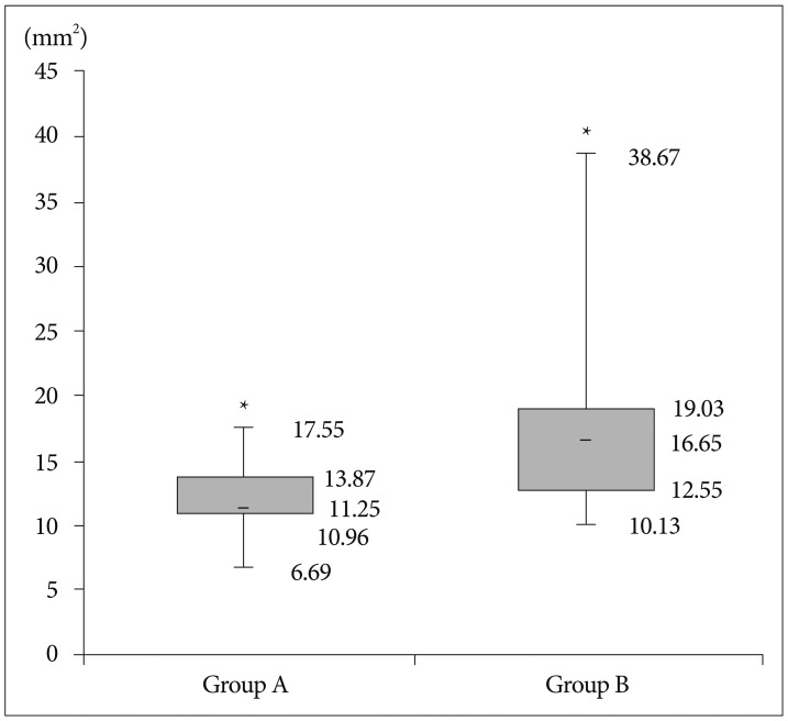
Of 213 patients with lumbar disc herniation encountered during the reference period, 35 patients who were followed up for 1 year after PELD were enrolled in this study. The patients were categorized by the period and technique of operation : group A, the first 15 cases, who underwent by the 'in-and-out' technique; group B, the next 20 cases, who underwent by the 'in-and-out-and-in' technique. The operation time, failure rate, blood loss, complication rate, re-herniation rate, the Visual Analogue Scale (VAS) for back and leg were checked. The alteration of dural sac cross-sectional area (DSCSA) between the preoperative and the postoperative MRI was checked.
RESULTS
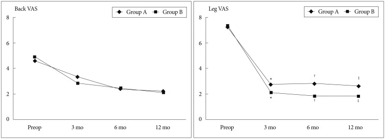
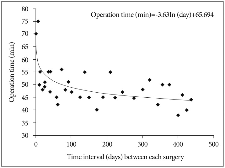
Operative time was rapidly reduced in the early phase, and then tapered to a steady state for the 35 cases receiving the PELD. After surgery, VAS scores for the back and leg were decreased significantly in both groups. Complications occurred in 2 patients in group A and 2 patients in group B. Between the two groups, there were significant differences in operative time, improvement of leg VAS, and expansion of DSCSA.
CONCLUSION
PELD learning curve seems to be acceptable with sufficient preparation. However, because of their high tendency to delayed operation time, operation failure, and re-herniation, caution should be exercised at the early phase of the procedure.
MeSH Terms
Figure
Reference
-
1. Chae KH, Ju CI, Lee SM, Kim BW, Kim SY, Kim HS. Strategies for noncontained lumbar disc herniation by an endoscopic approach : transforaminal suprapedicular approach, semi-rigid flexible curved probe, and 3-dimensional reconstruction CT with discogram. J Korean Neurosurg Soc. 2009; 46:312–316. PMID: 19893718.
Article2. Cho JY, Lee SH, Lee HY. Prevention of development of postoperative dysesthesia in transforaminal percutaneous endoscopic lumbar discectomy for intracanalicular lumbar disc herniation : floating retraction technique. Minim Invasive Neurosurg. 2011; 54:214–218. PMID: 22287030.
Article3. Choi G, Kang HY, Modi HN, Prada N, Nicolau RJ, Joh JY, et al. Risk of developing seizure after percutaneous endoscopic lumbar discectomy. J Spinal Disord Tech. 2011; 24:83–92. PMID: 20625320.
Article4. Choi G, Modi HN, Prada N, Ahn TJ, Myung SH, Gang MS, et al. Clinical results of XMR-assisted percutaneous transforaminal endoscopic lumbar discectomy. J Orthop Surg Res. 2013; 8:14. PMID: 23705685.
Article5. Choi I, Ahn JO, So WS, Lee SJ, Choi IJ, Kim H. Exiting root injury in transforaminal endoscopic discectomy : preoperative image considerations for safety. Eur Spine J. 2013; 22:2481–2487. PMID: 23754603.
Article6. Choi KC, Kim JS, Ryu KS, Kang BU, Ahn Y, Lee SH. Percutaneous endoscopic lumbar discectomy for L5-S1 disc herniation : transforaminal versus interlaminar approach. Pain Physician. 2013; 16:547–556. PMID: 24284840.7. Han IH, Choi BK, Cho WH, Nam KH. The obturator guiding technique in percutaneous endoscopic lumbar discectomy. J Korean Neurosurg Soc. 2012; 51:182–186. PMID: 22639720.
Article8. Hirano Y, Mizuno J, Takeda M, Itoh Y, Matsuoka H, Watanabe K. Percutaneous endoscopic lumbar discectomy - early clinical experience. Neurol Med Chir (Tokyo). 2012; 52:625–630. PMID: 23006872.
Article9. Hsu HT, Chang SJ, Yang SS, Chai CL. Learning curve of full-endoscopic lumbar discectomy. Eur Spine J. 2013; 22:727–733. PMID: 23076645.
Article10. Jasper GP, Francisco GM, Telfeian AE. Endoscopic transforaminal discectomy for an extruded lumbar disc herniation. Pain Physician. 2013; 16:E31–E35. PMID: 23340542.11. Joh JY, Choi G, Kong BJ, Park HS, Lee SH, Chang SH. Comparative study of neck pain in relation to increase of cervical epidural pressure during percutaneous endoscopic lumbar discectomy. Spine (Phila Pa 1976). 2009; 34:2033–2038. PMID: 19675511.
Article12. Kafadar A, Kahraman S, Akbörü M. Percutaneous endoscopic transforaminal lumbar discectomy : a critical appraisal. Minim Invasive Neurosurg. 2006; 49:74–79. PMID: 16708335.
Article13. Kang SH, Park SW. Symptomatic post-discectomy pseudocyst after endoscopic lumbar discectomy. J Korean Neurosurg Soc. 2011; 49:31–36. PMID: 21494360.
Article14. Kim HS, Ju CI, Kim SW, Kim JG. Endoscopic transforaminal suprapedicular approach in high grade inferior migrated lumbar disc herniation. J Korean Neurosurg Soc. 2009; 45:67–73. PMID: 19274114.
Article15. Kim HS, Park JY. Comparative assessment of different percutaneous endoscopic interlaminar lumbar discectomy (PEID) techniques. Pain Physician. 2013; 16:359–367. PMID: 23877452.16. Lee DY, Ahn Y, Lee SH. Percutaneous endoscopic lumbar discectomy for adolescent lumbar disc herniation : surgical outcomes in 46 consecutive patients. Mt Sinai J Med. 2006; 73:864–870. PMID: 17117312.17. Lee DY, Lee SH. Learning curve for percutaneous endoscopic lumbar discectomy. Neurol Med Chir (Tokyo). 2008; 48:383–388. discussion 388-389PMID: 18812679.
Article18. Lee S, Kim SK, Lee SH, Kim WJ, Choi WC, Choi G, et al. Percutaneous endoscopic lumbar discectomy for migrated disc herniation : classification of disc migration and surgical approaches. Eur Spine J. 2007; 16:431–437. PMID: 16972067.
Article19. Lohman CM, Tallroth K, Kettunen JA, Lindgren KA. Comparison of radiologic signs and clinical symptoms of spinal stenosis. Spine (Phila Pa 1976). 2006; 31:1834–1840. PMID: 16845360.
Article20. Ruetten S, Komp M, Merk H, Godolias G. Use of newly developed instruments and endoscopes : full-endoscopic resection of lumbar disc herniations via the interlaminar and lateral transforaminal approach. J Neurosurg Spine. 2007; 6:521–530. PMID: 17561740.
Article21. Sairyo K, Egawa H, Matsuura T, Takahashi M, Higashino K, Sakai T, et al. State of the art : transforaminal approach for percutaneous endoscopic lumbar discectomy under local anesthesia. J Med Invest. 2014; 61:217–225. PMID: 25264038.
Article22. Sairyo K, Matsuura T, Higashino K, Sakai T, Takata Y, Goda Y, et al. Surgery related complications in percutaneous endoscopic lumbar discectomy under local anesthesia. J Med Invest. 2014; 61:264–269. PMID: 25264043.
Article23. Sencer A, Yorukoglu AG, Akcakaya MO, Aras Y, Aydoseli A, Boyali O, et al. Fully endoscopic interlaminar and transforaminal lumbar discectomy : short-term clinical results of 163 surgically treated patients. World Neurosurg. 2014; 82:884–890. PMID: 24907438.
Article24. Sirvanci M, Bhatia M, Ganiyusufoglu KA, Duran C, Tezer M, Ozturk C, et al. Degenerative lumbar spinal stenosis : correlation with Oswestry Disability Index and MR imaging. Eur Spine J. 2008; 17:679–685. PMID: 18324426.
Article25. Wang H, Huang B, Zheng W, Li C, Zhang Z, Wang J, et al. Comparison of early and late percutaneous endoscopic lumbar discectomy for lumbar disc herniation. Acta Neurochir (Wien). 2013; 155:1931–1936. PMID: 23975645.
Article26. Xin G, Shi-Sheng H, Hai-Long Z. Morphometric analysis of the YESS and TESSYS techniques of percutaneous transforaminal endoscopic lumbar discectomy. Clin Anat. 2013; 26:728–734. PMID: 23824995.
Article27. Yeung AT, Tsou PM. Posterolateral endoscopic excision for lumbar disc herniation : surgical technique, outcome, and complications in 307 consecutive cases. Spine (Phila Pa 1976). 2002; 27:722–731. PMID: 11923665.
- Full Text Links
- Actions
-
Cited
- CITED
-
- Close
- Share
- Similar articles
-
- Factors Affecting Learning Curve in Endoscopic Lumbar Discectomy using Interlaminar Approach
- Percutaneous Endoscopic Lumbar Discectomy (PELD)
- Transforaminal Endoscopic Thoracic Discectomy: Technical Review to Prevent Complications
- Clinical Results of Percutaneous Endoscopic Discectomy in Herniated Intervertebral disc of Lumbar Spine
- Clinical Analysis of the Automated Percutaneous Discectomy

