Diagnostic Efficacy of Contrast-Enhanced Ultrasound for Small Renal Masses
- Affiliations
-
- 1Department of Urology, Institute of Wonkwang Medical Science, Wonkwang University School of Medicine, Iksan, Korea. seraph@wku.ac.kr
- 2Department of Radiology, Institute of Wonkwang Medical Science, Wonkwang University School of Medicine, Iksan, Korea.
- KMID: 2069776
- DOI: http://doi.org/10.4111/kju.2014.55.9.587
Abstract
- PURPOSE
Ultrasound (US) is highly sensitive in the detection of renal masses. However, it may not be able to differentiate benign and malignant lesions in smaller masses. The purpose of this study was to determine the diagnostic efficacy of contrast-enhanced ultrasound (CEUS) for small renal masses.
MATERIALS AND METHODS
From January 2011 to December 2013, a total of 85 patients underwent CEUS for evaluation of renal masses. Of these patients, CEUS findings were retrospectively analyzed for small renal cell carcinoma (RCC) cases (n=38) and angiomyolipoma (AML) cases (n=11). The tumor echogenicity and enhancement patterns and degrees were evaluated. The diagnostic efficacy of CEUS in differentiating the two diseases was compared.
RESULTS
On CEUS, the findings of diffuse heterogeneous enhancement (observed in 78.9% of RCCs and 27.3% of AMLs, p=0.003), washout from hyperenhancement or iso-enhancement to hypoenhancement in late phase (73.7% of RCCs and 18.2% of AMLs, p=0.001), and perilesional rim-like enhancement (57.9% of RCCs and 9.1% of AMLs, p=0.006) were significantly different between AML and RCC cases. The corresponding sensitivity, specificity, positive predictive value, negative predictive value, and accuracy were 86.8% (33/38), 63.6% (7/11), 89.2% (33/37), 58.3% (7/12), and 81.6% (40/49), respectively.
CONCLUSIONS
Our results suggest that the characteristic CEUS features could have diagnostic value in the evaluation of small renal mass. CEUS showed a higher diagnostic efficacy than conventional US for differentiating RCC and AML.
MeSH Terms
-
Adult
Aged
Aged, 80 and over
Angiomyolipoma/*ultrasonography
Carcinoma, Renal Cell/*ultrasonography
Contrast Media/diagnostic use
Diagnosis, Differential
Female
Humans
Kidney Neoplasms/*ultrasonography
Male
Middle Aged
Reproducibility of Results
Retrospective Studies
Sensitivity and Specificity
Sulfur Hexafluoride/diagnostic use
Ultrasonography/*methods
Contrast Media
Sulfur Hexafluoride
Figure
-
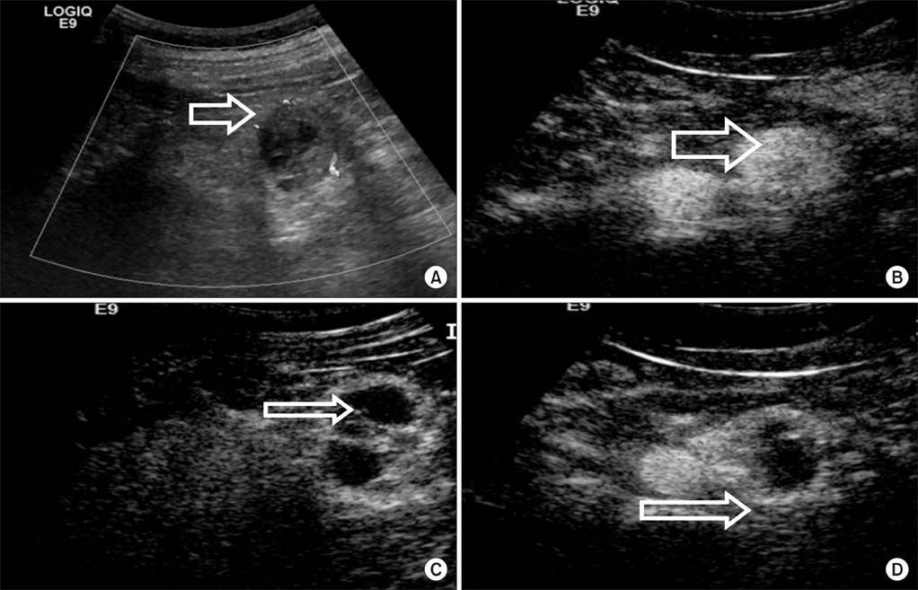
FIG. 1 A 42-year-old man with clear cell renal carcinoma. (A) Conventional ultrasonography demonstrated that a hypoechogenic mass with a diameter of 2.8 cm was located in the left lower kidney. (B) Contrast-enhanced ultrasound (CEUS) imaging of the arterial phase showed a diffuse, heterogeneous enhanced tumor. (C) CEUS imaging at the late phase showed washout of the tumor. (D) CEUS imaging at the late phase showed perilesional enhancement with slow centripetal fill in a rim-like pattern.

FIG. 2 A 56-year-old man with angiomyolipoma. (A) Conventional ultrasonography demonstrated that a hyperechogenic mass with a diameter of 2.3 cm was located in the left lower kidney. (B) Contrast-enhanced ultrasound imaging in the late phase showed a dot-like enhancement.
Reference
-
1. Forman HP, Middleton WD, Melson GL, McClennan BL. Hyperechoic renal cell carcinomas: increase in detection at US. Radiology. 1993; 188:431–434.2. Heidenreich A, Ravery V. European Society of Oncological Urology. Preoperative imaging in renal cell cancer. World J Urol. 2004; 22:307–315.3. Claudon M, Cosgrove D, Albrecht T, Bolondi L, Bosio M, Calliada F, et al. Guidelines and good clinical practice recommendations for contrast enhanced ultrasound (CEUS) - update 2008. Ultraschall Med. 2008; 29:28–44.4. Xu ZF, Xu HX, Xie XY, Liu GJ, Zheng YL, Liang JY, et al. Renal cell carcinoma: real-time contrast-enhanced ultrasound findings. Abdom Imaging. 2010; 35:750–756.5. Lu Q, Wang W, Huang B, Li C, Li C. Minimal fat renal angiomyolipoma: the initial study with contrast-enhanced ultrasonography. Ultrasound Med Biol. 2012; 38:1896–1901.6. Nilsson A. Contrast-enhanced ultrasound of the kidneys. Eur Radiol. 2004; 14:Suppl 8. P104–P109.7. Ascenti G, Gaeta M, Magno C, Mazziotti S, Blandino A, Melloni D, et al. Contrast-enhanced second-harmonic sonography in the detection of pseudocapsule in renal cell carcinoma. AJR Am J Roentgenol. 2004; 182:1525–1530.8. Zhou X, Yan F, Luo Y, Peng YL, Parajuly SS, Wen XR, et al. Characterization and diagnostic confidence of contrast-enhanced ultrasound for solid renal tumors. Ultrasound Med Biol. 2011; 37:845–853.9. Zhang J, Lefkowitz RA, Ishill NM, Wang L, Moskowitz CS, Russo P, et al. Solid renal cortical tumors: differentiation with CT. Radiology. 2007; 244:494–504.10. Russo P. Renal cell carcinoma: presentation, staging, and surgical treatment. Semin Oncol. 2000; 27:160–176.11. Pavlica P, Derchi L, Martorana G, Brunocilla E, Bertaccini A, Manferrari F, et al. Renal cell carcinoma imaging. Eur Urol Suppl. 2006; 5:580–592.12. Jinzaki M, Tanimoto A, Narimatsu Y, Ohkuma K, Kurata T, Shinmoto H, et al. Angiomyolipoma: imaging findings in lesions with minimal fat. Radiology. 1997; 205:497–502.13. Quaia E, Palumbo A, Rossi S, Degobbis F, Cernic S, Tona G, et al. Comparison of visual and quantitative analysis for characterization of insonated liver tumors after microbubble contrast injection. AJR Am J Roentgenol. 2006; 186:1560–1570.14. Quaia E, Calliada F, Bertolotto M, Rossi S, Garioni L, Rosa L, et al. Characterization of focal liver lesions with contrast-specific US modes and a sulfur hexafluoride-filled microbubble contrast agent: diagnostic performance and confidence. Radiology. 2004; 232:420–430.15. Xu ZF, Xu HX, Xie XY, Liu GJ, Zheng YL, Lu MD. Renal cell carcinoma and renal angiomyolipoma: differential diagnosis with real-time contrast-enhanced ultrasonography. J Ultrasound Med. 2010; 29:709–717.16. Jinzaki M, Tanimoto A, Mukai M, Ikeda E, Kobayashi S, Yuasa Y, et al. Double-phase helical CT of small renal parenchymal neoplasms: correlation with pathologic findings and tumor angiogenesis. J Comput Assist Tomogr. 2000; 24:835–842.17. Pickhardt PJ, Lonergan GJ, Davis CJ Jr, Kashitani N, Wagner BJ. From the archives of the AFIP. Infiltrative renal lesions: radiologic-pathologic correlation. Armed Forces Institute of Pathology. Radiographics. 2000; 20:215–243.18. Setola SV, Catalano O, Sandomenico F, Siani A. Contrast-enhanced sonography of the kidney. Abdom Imaging. 2007; 32:21–28.
- Full Text Links
-
- Actions
-
Cited
- CITED
-
- Close
- Share
-
- Similar articles
-
- New Ultrasonographic Techniques to Differentiate Hepatic Masses: Contrast-Enhanced Ultrasound and Super-Resolution Ultrasound
- Multislice computed tomography/contrast-enhanced ultrasound image fusion as a tool for evaluating unclear renal cysts
- MR Finding of Primary Renal Lymphoma: A Case Report
- Clinical use of contrast-enhanced ultrasound beyond the liver: a focus on renal, splenic, and pancreatic applications
- Assessment of Cystic Renal Masses Based on Bosniak Classification: Comparison of CT, Contrast-enhanced US, and MR Imaging
