Two Cases of Primary Osteolytic Intraosseous Meningioma of the Skull Metastasizing to Whole Skull and the Spine
- Affiliations
-
- 1Department of Neurosurgery, Chonnam National University Research Institute of Medical Sciences, Chonnam National University Hwasun Hospital & Medical School, Gwangju, Korea. jung-ty@chonnam.ac.kr
- KMID: 2018189
- DOI: http://doi.org/10.3340/jkns.2012.51.3.151
Abstract
- We report here two cases of primary intraosseous meningioma with aggressive behavior. A 68-year-old man presented with a one year history of a soft, enlarging mass in the right parietal region. Magnetic resonance image (MRI) revealed a 6 cm sized, heterogeneously-enhancing, bony expansile mass in the right parietal bone, and computed tomograph (CT) showed a bony, destructive lesion. The tumor, including the surrounding normal bone, was totally resected. Dural invasion was not apparent. Diagnosis was atypical meningioma, which extensively metastasized within the skull one year later. A 74-year-old woman presented with a 5-month history of a soft mass on the left frontal area. MRI revealed a 4 cm sized, multilobulated, strongly-enhancing lesion on the left frontal bone, and CT showed a destructive lesion. The mass was adhered tightly to the scalp and dura mater. The lesion was totally removed. Biopsy showed a papillary meningioma. The patient refused adjuvant radiation therapy and later underwent two reoperations for recurred lesions, at 19 and at 45 months postoperative. The patient experienced back pain 5 years later, and MRI showed an osteolytic lesion on the 11th thoracic vertebra. After her operation, a metastatic papillary meningioma was diagnosed. These osteolytic intraosseous meningiomas had atypical/malignant pathologies, which metastasized to whole skull and the spine.
Keyword
MeSH Terms
Figure
-
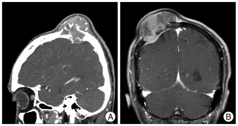
Fig. 1 Case 1 : Preoperative radiologic findings. A : CT image shows a bony, destructive lesion, in the right parietal bone abutting the scalp and dura. B : Coronal MRI revealing a heterogeneously-enhancing, bony, expansile mass in the right parietal bone, extending into intracranial epidural space.

Fig. 2 Case 1 : Pathologic findings (atypical meningioma). A : Cellular whorl formation (H&E, original magnification ×100). B : Immunopositive for epithelial membrane antigen (original magnification ×400). C : Frequent mitosis, (H&E, original magnification ×400). D : Necrosis (H&E, original magnification ×100)
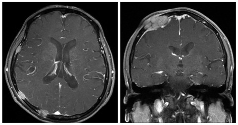
Fig. 3 Case 1 : Follow-up radiologic findings. One year later, follow-up brain MRI shows multiple variable sized nodules and masses in whole skull, causing epidural and subgaleal extraosseous mass formations.

Fig. 4 Case 2 : Preoperative radiologic findings. A : CT image shows a bony, destructive lesion in the left frontal bone. B : MRI revealing a 4-centimeter, multilobulated, strong-enhancing mass on the left frontal bone, with thickening of adjacent scalp and meninges.
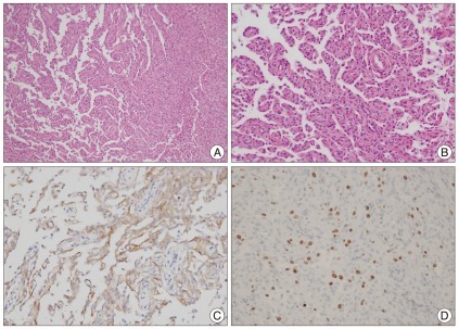
Fig. 5 Case 2 : Pathologic findings (papillary meningioma). A : Papillary structures mixed with meningothelial sheets (H&E, original magnification ×100). B : Tumor cells in papillary pattern (original magnification ×200). C : Immunopositive for epithelial membrane antigen (original magnification ×200). D : Three percent of Ki-67 labeling index (H&E, original magnification ×100).
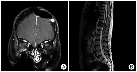
Fig. 6 Case 2 : Follow-up radiologic findings. A : Follow-up MRI demonstrates a 1 cm, homogenously-enhancing, recurred lesion on the skull, adjacent to the previous lesion. B : Spinal MRI shows the osteolytic lesion on the body and posterior element of the 11th thoracic vertebra.
Cited by 2 articles
-
Extracranial Extension of Intracranial Atypical Meningioma En Plaque with Osteoblastic Change of the Skull
Se Youn Jang, Choong Hyun Kim, Jin Hwan Cheong, Jae Min Kim
J Korean Neurosurg Soc. 2014;55(4):205-207. doi: 10.3340/jkns.2014.55.4.205.Primary Osteolytic Intraosseous Atypical Meningioma with Soft Tissue and Dural Invasion: Report of a Case and Review of Literatures
Jung-Ho Yun, Sang-Koo Lee
J Korean Neurosurg Soc. 2014;56(6):509-512. doi: 10.3340/jkns.2014.56.6.509.
Reference
-
1. Agrawal V, Ludwig N, Agrawal A, Bulsara KR. Intraosseous intracranial meningioma. AJNR Am J Neuroradiol. 2007; 28:314–315. PMID: 17297003.2. Arana E, Menor F, Lloret RM. Intraosseous meningioma. J Neurosurg. 1996; 85:362–363. PMID: 8755774.3. Crawford TS, Kleinschmidt-DeMasters BK, Lillehei KO. Primary intraosseous meningioma. Case report. J Neurosurg. 1995; 83:912–915. PMID: 7472564.4. Daffner RH, Yakulis R, Maroon JC. Intraosseous meningioma. Skeletal Radiol. 1998; 27:108–111. PMID: 9526778.
Article5. Devi B, Bhat D, Madhusudhan H, Santhosh V, Shankar S. Primary intraosseous meningioma of orbit and anterior cranial fossa : a case report and literature review. Australas Radiol. 2001; 45:211–214. PMID: 11380366.
Article6. Elder JB, Atkinson R, Zee CS, Chen TC. Primary intraosseous meningioma. Neurosurg Focus. 2007; 23:E13. PMID: 17961037.
Article8. Inagaki K, Otsuka F, Matsui T, Ogura T, Makino H. Effect of etidronate on intraosseous meningioma. Endocr J. 2004; 51:389–390. PMID: 15256788.
Article9. Jayaraj K, Martinez S, Freeman A, Lyles KW. Intraosseous meningioma--a mimicry of Paget's disease? J Bone Miner Res. 2001; 16:1154–1156. PMID: 11393793.10. Lang FF, Macdonald OK, Fuller GN, DeMonte F. Primary extradural meningiomas : a report on nine cases and review of the literature from the era of computerized tomography scanning. J Neurosurg. 2000; 93:940–950. PMID: 11117866.
Article11. Lapresle J, Netsky MG, Zimmerman HM. [The pathology of meningiomas; a study of 121 cases]. Am J Pathol. 1952; 28:757–791. PMID: 12976523.12. Lell M, Tudor C, Aigner T, Kessler P. Primary intraosseous meningioma of the mandible : CT and MR imaging features. AJNR Am J Neuroradiol. 2007; 28:129–131. PMID: 17213439.13. Marwah N, Gupta S, Marwah S, Singh S, Kalra R, Arora B. Primary intraosseous meningioma. Indian J Pathol Microbiol. 2008; 51:51–52. PMID: 18417855.
Article14. Partington MD, Scheithauer BW, Piepgras DG. Carcinoembryonic antigen production associated with an osteolytic meningioma. Case report. J Neurosurg. 1995; 82:489–492. PMID: 7861230.
Article15. Politi M, Romeike BF, Papanagiotou P, Nabhan A, Struffert T, Feiden W, et al. Intraosseous hemangioma of the skull with dural tail sign : radiologic features with pathologic correlation. AJNR Am J Neuroradiol. 2005; 26:2049–2052. PMID: 16155158.16. Shuangshoti S. Al-Mefty O, editor. Primary meningiomas outside the central nervous system. Meningiomas. 1991. New York: Raven Press;p. 107–128.
- Full Text Links
-
- Actions
-
Cited
- CITED
-
- Close
- Share
-
- Similar articles
-
- Primary Intraosseous Osteolytic Meningioma of the Skull Mimicking Scalp Mass: A Case Report and Review of Literature
- Primary Intraosseous Calvarial Meningioma: A Case Report
- Intradiploic Meningioma Presenting as on Osteolytic Skull Lesion Case Report
- Primary Osteolytic Intraosseous Atypical Meningioma with Soft Tissue and Dural Invasion: Report of a Case and Review of Literatures
- Ewing's Sarcoma Mimicking a Meningioma in Radiological Findings: A Case Report
