Clin Exp Otorhinolaryngol.
2009 Jun;2(2):78-84. 10.3342/ceo.2009.2.2.78.
Clinical Efficacy of Primary Tumor Volume Measurements: Comparison of Different Primary Sites
- Affiliations
-
- 1Department of Otolaryngology-Head and Neck Surgery, Korea University College of Medicine, Seoul, Korea. kyjung@kumc.or.kr
- 2Department of Radiology, Korea University College of Medicine, Seoul, Korea.
- KMID: 2007189
- DOI: http://doi.org/10.3342/ceo.2009.2.2.78
Abstract
OBJECTIVES
The purpose of study was to determine the clinical efficacy of primary tumor volume measurements of different primary sites in the oropharynx compared to the oral cavity.
METHODS
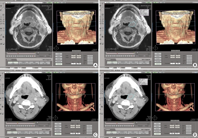
A retrospective analysis of 85 patients with oral cavity or oropharynx cancer. The tumor area was manually outlined from axial magnetic resonance (MR) series. The software calculated the tumor volumes, automatically. The values of the primary tumor volumes were then subdivided into separate groups (< or =3,500 mm3, >3,500 mm3).
RESULTS
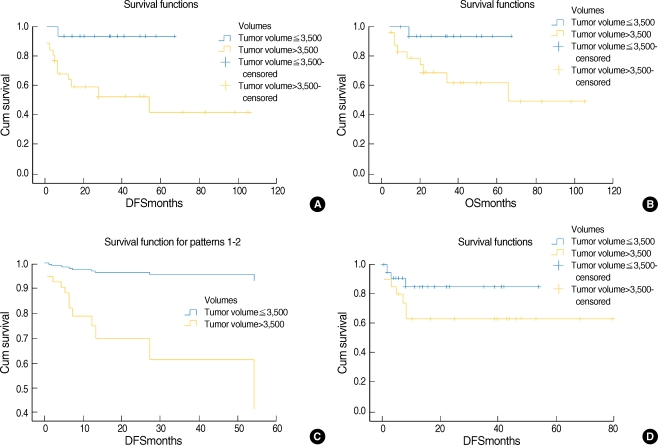
The prognostic indicators were the cT and cN (oral cavity); age, primary site, cT, cN, and primary tumor volume (oropharynx) on the univariate analysis. There was no significant prognostic factor for oral cavity cancer on the multivariate analysis. Primary site, cN, and primary tumor volume were independent prognostic indicators for oropharynx cancer by multivariate analysis.
CONCLUSION
Primary tumor volume measurement is a reliable way to stratify outcome, and make up for the weak points in the American Joint Committee on Cancer staging system with oropharynx cancer.
MeSH Terms
Figure
Reference
-
1. Kimura Y, Sumi M, Ichikawa Y, Kawai Y, Nakamura T. Volumetric MR imaging of oral, maxillary sinus, oropharyngeal, and hypopharyngeal cancers: correlation between tumor volume and lymph node metastasis. AJNR Am J Neuroradiol. 2005; 10. 26(9):2384–2389. PMID: 16219850.2. Le Tourneau C, Velten M, Jung GM, Bronner G, Flesch H, Borel C. Prognostic indicators for survival in head and neck squamous cell carcinomas: analysis of a series of 621 cases. Head Neck. 2005; 9. 27(9):801–808. PMID: 16086415.
Article3. Mancuso AA, Mukherji SK, Schmalfuss I, Mendenhall W, Parsons J, Pameijer F, et al. Preradiotherapy computed tomography as a predictor of local control in supraglottic carcinoma. J Clin Oncol. 1999; 2. 17(2):631–637. PMID: 10080608.
Article4. Lo SM, Venkatesan V, Matthews TW, Rogers J. Tumour volume: implications in T2/T3 glottic/supraglottic squamous cell carcinoma. J Otolaryngol. 1998; 10. 27(5):247–251. PMID: 9800621.5. Chao KS, Ozyigit G, Blanco AI, Thorstad WL, Deasy JO, Haughey BH, et al. Intensity-modulated radiation therapy for oropharyngeal carcinoma: impact of tumor volume. Int J Radiat Oncol Biol Phys. 2004; 5. 01. 59(1):43–50. PMID: 15093897.
Article6. Doweck I, Denys D, Robbins KT. Tumor volume predicts outcome for advanced head and neck cancer treated with targeted chemoradiotherapy. Laryngoscope. 2002; 10. 112(10):1742–1749. PMID: 12368607.
Article7. Johnson CR, Thames HD, Huang DT, Schmidt-Ullrich RK. The tumor volume and clonogen number relationship: tumor control predictions based upon tumor volume estimates derived from computed tomography. Int J Radiat Oncol Biol Phys. 1995; 9. 30. 33(2):281–287. PMID: 7673015.8. Johnson CR, Khandelwal SR, Schmidt-Ullrich RK, Ravalese J 3rd, Wazer DE. The influence of quantitative tumor volume measurements on local control in advanced head and neck cancer using concomitant boost accelerated superfractionated irradiation. Int J Radiat Oncol Biol Phys. 1995; 6. 15. 32(3):635–641. PMID: 7790249.
Article9. Chang CC, Chen MK, Liu MT, Wu HK. The effect of primary tumor volumes in advanced T-staged nasopharyngeal tumors. Head Neck. 2002; 10. 24(10):940–946. PMID: 12369073.
Article10. Weiss E, Hess CF. The impact of gross tumor volume (GTV) and clinical target volume (CTV) definition on the total accuracy in radiotherapy theoretical aspects and practical experiences. Strahlenther Onkol. 2003; 1. 179(1):21–30. PMID: 12540981.11. Gordon AR, Loevner LA, Shukla-Dave A, Redfern RO, Sonners AI, Kilger AM, et al. Intraobserver variability in the MR determination of tumor volume in squamous cell carcinoma of the pharynx. AJNR Am J Neuroradiol. 2004; Jun–Jul. 25(6):1092–1098. PMID: 15205156.
- Full Text Links
- Actions
-
Cited
- CITED
-
- Close
- Share
- Similar articles
-
- Clinical Analysis of Small Bowel Tumor
- A Case of Primary Malignant Mixed Mullerian Tumor of the Pelvic Peritoneum
- Circulating Tumor Cell Number Is Associated with Primary Tumor Volume in Patients with Lung Adenocarcinoma
- Cytologic Features and Distribution of Primary site of Malignant Cells in Body Fluids
- Clinical Characteristics and Prognostic Factors of Metastatic Tumor of Unknown Primary


