J Korean Soc Spine Surg.
2003 Jun;10(2):154-162. 10.4184/jkss.2003.10.2.154.
Treatment of Degenerative Lumbar Stenosis with Minimal Decompression
- Affiliations
-
- 1Department of Orthopedic Surgery, Samsun Hospital, Busan, Korea. c3k10127@korea.com
- KMID: 2003213
- DOI: http://doi.org/10.4184/jkss.2003.10.2.154
Abstract
- STUDY DESIGN: A retrospective study
OBJECTIVES
In the operative treatment of lumbar spinal stenosis, the wide decompression and fusion method has many problems, such as a long operation time, large blood loss and the long time required to achieve solid fusion. As a solution to these problems, a minimal decompression method was been performed, which minimizes the resection of laminae and facet joints. SUMMARY OF LITERATURE REVIEW: In the operative therapy for lumbar spinal stenosis, favorable results can be obtained by simple decompression.
MATERIALS AND METHODS
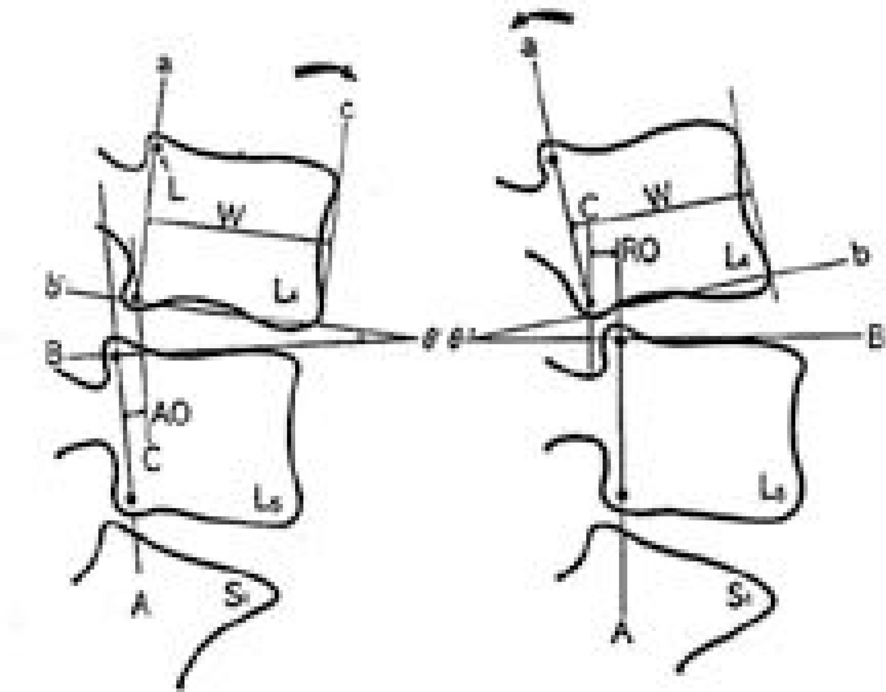
42 cases of degenerative lumbar stenosis, with neither segmental instability nor spondylolisthesis, underwent a minimal decompressive surgery, without instrumentation. The mean operation time and amount of blood loss were analyzed, and the clinical results evaluated according to Kim's criteria and the postoperative segmental instability by the Dupuis method. The average follow-up period was 70 months.
RESULTS
Transfusions were not required in all cases. The mean operative times were 1hour 5minutes and 1hour 46minutes in the one and two segment decompressions, respectively. The clinical results, according to Kim's criteria, were excellent in 24 cases and good in 12. There was no dynamic instability in the radiographs at the last follow-up.
CONCLUSIONS
With the degenerative lumbar stenosis, without segmental instability or spondylolisthesis, minimal decompression was an effective surgical method.
Keyword
MeSH Terms
Figure
Cited by 1 articles
-
Minimally Invasive Microscopic Decompression with Tubular Retractor System in Lumbar Spinal Stenosis - Results Comparing with Open Microscopic Decompression -
Jae Ho Jang, Jae Do Kim, Sang Won Cha
J Korean Soc Spine Surg. 2007;14(2):79-86. doi: 10.4184/jkss.2007.14.2.79.
Reference
-
1). Abmui K., Panjabi MM., Kramer KM. Biomechani -cal evaluation of lumbar spinals instability after graded facetectomies. Spine,. 15:1142–1147. 1990.2). Benz RJ., Garfin SR. Current techniques of decom -pression of the lumbar spine. Clin Orthop,. 384:75–81. 2001.3). Crock H., Crock M. A technique for decompression of the lumbar spinal canal. Neuro-Orthopedics,. 5:96–99. 1988.4). Dupuis PR., Yong-hing K., Cassidy JD. Radiologic diagnosis of degenerative lumbar spinal instability. Spine,. 10(3):262–76. 1985.
Article5). Frymoyer JW., Hanley EN., Howe J. A Comparision of radiographic findings in fusion and nonfusion patients ten or more years following lumbar disk surgery. Spine,. 4(5):435–440. 1979.6). Grob D., Humke T., Dvorak J. Degenerative lumbar spinal stenosis. Decompression with and without arthrodesis. J Bone Joint Surg,. 77(A7):1036–41. 1995.
Article7). Jeffrey M Spivak. Current Concepts of Review-Degener -ative Lumbar Spinal Stenosis. J Bone Joint Surg,. 80(A):1053–66. 1998.8). Kim NH., Seo IK. The Effect of Anterior Interbody Fusion in Lumbar Hernated Nucleus Pulposus. J of Kor Orthop Assoc,. 21(2):202–10. 1986.9). Leong JCY., Grange WJ., Fang D. Long term results of lumbar intervertebral disc prolapse. Spine,. 8:793–799. 1983.10). Shim DM., Kim SS., Han HJ., Lee BC., Shin JH. Selective spinal root block method used for testing lumbar spinal disease. J Korean Spine Surg,. 1:293–299. 1996.11). Shin BJ., Shin YS., Kwon H., Yim SJ., Kim DS., Choi CU. The results of treatment of multilevel spinal stenosis. J Kor Spine Surg,. 3:161–168. 1996.12). Suk SI. Spinal surgery. Newest medicine Co;p. 223–227. 1997.
Article13). S. Terry Canale. International Edition of Campbell's Operative Orthopaedics. 10th ed.Philadelphia: Mosby;p. 1959–1961. 2003.14). Thomas J. Gill and Michael D. Mason. Assessment of neuroforaminal decompression in degenerative spinal stenosis. Clin Orthop,. 348:135–139. 1998.15). White AA III., Panjabi MM. Clinical biomechanics of the spine. 2nd ed.Philadelphia: JB Linppincott Co;1990.
- Full Text Links
- Actions
-
Cited
- CITED
-
- Close
- Share
- Similar articles
-
- Result of Pedicle Screw Fixation in Lumbar Stenosis with: A Comparison of Degenerative Type Lumbar Stenosis with Spondylolisthetic type Lumbar Stenosis
- A Clinical Study on the Causes of the Nerve Entrapment in the Degenerative Spondylolisthesis
- A Comparison of Clinical Outcomes between Decompressive Lumbar Laminectomy Alone and with Arthrodesis in Degenerative Lumbar Spinal Stenosis
- Cotrel - Dubousset Pedicle Screw Fixation After Posterior Decompression of Lumbar Spinal Stenosis
- Results of Decompression Alone in Patients with Lumbar Spinal Stenosis and Degenerative Spondylolisthesis: A Minimum 5-Year Follow-up


