J Breast Cancer.
2013 Dec;16(4):442-446. 10.4048/jbc.2013.16.4.442.
Random Synchronous Malignancy in Male Breast: A Case Report
- Affiliations
-
- 1Department of Nuclear Medicine & PET CT, Amrita Institute of Medical Sciences (Amrita Vishwa Vidyapeetham), Cochin, Kerala, India. drmanjitsarma@gmail.com
- KMID: 1976384
- DOI: http://doi.org/10.4048/jbc.2013.16.4.442
Abstract
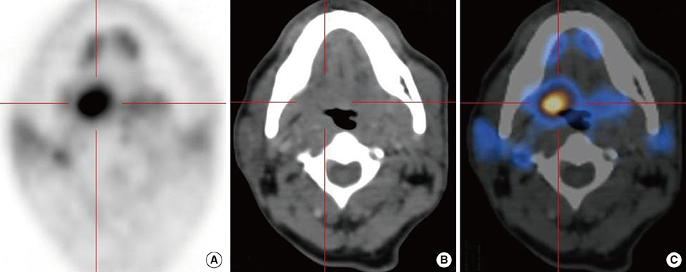
- We report here a case of a random synchronous male breast malignancy in a patient with a known base of tongue malignancy that was incidentally detected on a whole body 18-fluorine deoxyglucose positron emission tomography and computed tomography (18F-FDG PET/CT). Patient was referred to us for PET/CT staging and radiotherapy planning for a poorly differentiated squamous cell carcinoma of base of tongue. Histopathologically, the incidentally detected breast lesion was proven to be an invasive ductal carcinoma. 18F-FDG PET/CT being a whole body imaging modality is known to detect a considerable number of synchronous primaries. Synchronous malignancies in the head and neck area and the upper aerodigestive tract are well established. However, synchronous malignancy in male breast is reportedly uncommon. Our case is unique for the fact that a random synchronous dual malignancy of base of tongue and breast in a male patient was detected during a whole body 18F-FDG PET/CT imaging.
Keyword
MeSH Terms
-
Breast Neoplasms
Breast*
Carcinoma, Ductal
Carcinoma, Squamous Cell
Deoxyglucose
Fluorodeoxyglucose F18
Head
Humans
Male*
Neck
Neoplasms, Multiple Primary
Positron-Emission Tomography
Positron-Emission Tomography and Computed Tomography
Radiotherapy
Tongue
Tongue Neoplasms
Whole Body Imaging
Deoxyglucose
Fluorodeoxyglucose F18
Figure
Reference
-
1. Gluckman JL, Crissman JD, Donegan JO. Multicentric squamous-cell carcinoma of the upper aerodigestive tract. Head Neck Surg. 1980; 3:90–96.
Article2. Rennemo E, Zätterström U, Boysen M. Synchronous second primary tumors in 2,016 head and neck cancer patients: role of symptom-directed panendoscopy. Laryngoscope. 2011; 121:304–309.
Article3. Slaughter DP, Southwick HW, Smejkal W. Field cancerization in oral stratified squamous epithelium: clinical implications of multicentric origin. Cancer. 1953; 6:963–968.
Article4. Yaman E, Ozturk B, Coskun U, Buyukberber S, Kaya AO, Yildiz R, et al. Synchronous bilateral breast cancer in an aged male patient. Onkologie. 2010; 33:255–258.
Article5. Sun WY, Lee KH, Lee HC, Ryu DH, Park JW, Yun HY, et al. Synchronous bilateral male breast cancer: a case report. J Breast Cancer. 2012; 15:248–251.
Article6. Dubashi B, Jain A, Srinivasan K, Surendrakumar V, Vivekanandam S. Chronic lymphocytic leukemia and breast cancer as synchronous primary in a male-a rare combination. Curr Oncol. 2011; 18:e101–e102.
Article7. Sordi E, Cagossi K, Lazzaretti MG, Gusolfino D, Artioli F, Santacroce G, et al. Rare case of male breast cancer and axillary lymphoma in the same patient: an unique case report. Case Rep Med. 2011; 2011:940803.
Article8. Strobel K, Haerle SK, Stoeckli SJ, Schrank M, Soyka JD, Veit-Haibach P, et al. Head and neck squamous cell carcinoma (HNSCC): detection of synchronous primaries with (18)F-FDG-PET/CT. Eur J Nucl Med Mol Imaging. 2009; 36:919–927.
Article9. Mesmoudi M, Mahfoud T, Ismaili N, Rami K, Kamouni M, Jroundi L, et al. A synchronous undifferentiated nasopharyngeal carcinoma and infiltrating ductal carcinoma of the breast successfully treated with induction chemotherapy followed by local control of both tumours: a case report. BMC Ear Nose Throat Disord. 2011; 11:6.
Article10. Demandante CG, Troyer DA, Miles TP. Multiple primary malignant neoplasms: case report and a comprehensive review of the literature. Am J Clin Oncol. 2003; 26:79–83.11. Warren S, Gates O. Multiple primary malignant tumors: a survey of the literature and a statistical study. Am J Cancer. 1932; 16:1358–1414.12. Moertel CG. Multiple primary malignant neoplasms: historical perspectives. Cancer. 1977; 40:4 Suppl. 1786–1792.
Article13. Tamura M, Shinagawa M, Funaki Y. Synchronous triple early cancers occurring in the stomach, colon and gallbladder. Asian J Surg. 2003; 26:46–48.
Article14. Meguerditchian AN, Falardeau M, Martin G. Male breast carcinoma. Can J Surg. 2002; 45:296–302.15. Goss PE, Reid C, Pintilie M, Lim R, Miller N. Male breast carcinoma: a review of 229 patients who presented to the Princess Margaret Hospital during 40 years: 1955-1996. Cancer. 1999; 85:629–639.
- Full Text Links
- Actions
-
Cited
- CITED
-
- Close
- Share
- Similar articles
-
- Male Patients with the Diagnoses of Synchronous Prostate and Breast Cancer
- Positive predictive value of additional synchronous breast lesions in whole-breast ultrasonography at the diagnosis of breast cancer: clinical and imaging factors
- Synchronous BI-RADS Category 3 Lesions on Preoperative Ultrasonography in Patients with Breast Cancer: Is Short-Term Follow-Up Appropriate?
- Myoid Hamartoma of the Breast with Synchronous Contralateral Breast Cancer: Report of a Case
- Synchronous Bilateral Male Breast Cancer: A Case Report


