J Breast Cancer.
2013 Dec;16(4):386-394. 10.4048/jbc.2013.16.4.386.
Effect Modification of Hormonal Therapy by p53 Status in Invasive Breast Cancer
- Affiliations
-
- 1Department of Surgery, Asan Medical Center, University of Ulsan College of Medicine, Seoul, Korea. asanbreastcenter@gmail.com
- 2Department of Biostatistics and Clinical Epidemiology, Asan Medical Center, University of Ulsan College of Medicine, Seoul, Korea.
- 3Department of Surgery, Seoul National University College of Medicine, Seoul, Korea.
- 4Department of Surgery, Keimyung University School of Medicine, Daegu, Korea.
- 5Department of Pathology, Asan Medical Center, University of Ulsan College of Medicine, Seoul, Korea.
- 6Department of Oncology, Asan Medical Center, University of Ulsan College of Medicine, Seoul, Korea.
- KMID: 1976376
- DOI: http://doi.org/10.4048/jbc.2013.16.4.386
Abstract
- PURPOSE
We aimed to confirm the prognostic and predictive value of p53 expression, particularly in invasive breast cancer patients, according to immunohistochemical hormone receptor (HR) and human epidermal growth factor receptor 2 (HER2) status.
METHODS
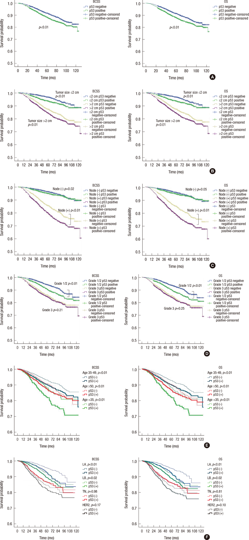
Immunohistochemical data for p53, estrogen receptor, progesterone receptor, and HER2 expression from a total of 15,598 patients were retrospectively retrieved from the web-based database of the Korean Breast Cancer Society. Overall survival (OS) and breast cancer-specific survival (BCSS) were calculated and compared using the Kaplan-Meier method and log-rank test, respectively. Multivariate analyses were performed using a stratified Cox proportional hazard regression model. A model evaluating interactions between p53 expression and both hormonal therapy and chemotherapy was used to determine the treatment benefit from both modalities.
RESULTS
The prognostic value of p53 for OS and BCSS was most significant in the HR+/HER2- subgroup, with hazard ratios of 1.44 (95% confidence interval [CI], 1.08-1.93) and 1.47 (95% CI, 1.09-1.99), respectively. The p53 overexpression hazard ratios were of borderline significance for the HR+/HER2+ subgroup and were not significant for the HR-/HER2+ and HR-/HER2- subgroups. The model with interaction terms revealed that hormonal therapy significantly interacts with p53 status (p=0.002 and p=0.007 for OS and BCSS, respectively), suggesting an insignificant prognostic value for p53 status (p=0.268 and p=0.296 for OS and BCSS, respectively). An interaction between chemotherapy and p53 status was not found in this model.
CONCLUSION
p53 overexpression has independent prognostic value, particularly in cases of HR+/HER2- invasive breast cancer, which may be due to effect modification of hormonal therapy dependent on p53 status.
MeSH Terms
Figure
Reference
-
1. Silvestrini R, Daidone MG, Benini E, Faranda A, Tomasic G, Boracchi P, et al. Validation of p53 accumulation as a predictor of distant metastasis at 10 years of follow-up in 1400 node-negative breast cancers. Clin Cancer Res. 1996; 2:2007–2013.2. Krajewski S, Krajewska M, Turner BC, Pratt C, Howard B, Zapata JM, et al. Prognostic significance of apoptosis regulators in breast cancer. Endocr Relat Cancer. 1999; 6:29–40.
Article3. Malamou-Mitsi V, Gogas H, Dafni U, Bourli A, Fillipidis T, Sotiropoulou M, et al. Evaluation of the prognostic and predictive value of p53 and Bcl-2 in breast cancer patients participating in a randomized study with dose-dense sequential adjuvant chemotherapy. Ann Oncol. 2006; 17:1504–1511.
Article4. Marks JR, Humphrey PA, Wu K, Berry D, Bandarenko N, Kerns BJ, et al. Overexpression of p53 and HER-2/neu proteins as prognostic markers in early stage breast cancer. Ann Surg. 1994; 219:332–341.
Article5. Han JS, Cao D, Molberg KH, Sarode VR, Rao R, Sutton LM, et al. Hormone receptor status rather than HER2 status is significantly associated with increased Ki-67 and p53 expression in triple-negative breast carcinomas, and high expression of Ki-67 but not p53 is significantly associated with axillary nodal metastasis in triple-negative and high-grade non-triple-negative breast carcinomas. Am J Clin Pathol. 2011; 135:230–237.
Article6. Haerslev T, Jacobsen GK. An immunohistochemical study of p53 with correlations to histopathological parameters, c-erbB-2, proliferating cell nuclear antigen, and prognosis. Hum Pathol. 1995; 26:295–301.
Article7. Barbareschi M, Caffo O, Veronese S, Leek RD, Fina P, Fox S, et al. Bcl-2 and p53 expression in node-negative breast carcinoma: a study with long-term follow-up. Hum Pathol. 1996; 27:1149–1155.
Article8. Lê MG, Mathieu MC, Douc-Rasy S, Le Bihan ML, Adb El All H, Spielmann M, et al. c-myc, p53 and bcl-2, apoptosis-related genes in infiltrating breast carcinomas: evidence of a link between bcl-2 protein over-expression and a lower risk of metastasis and death in operable patients. Int J Cancer. 1999; 84:562–567.9. Reed W, Hannisdal E, Boehler PJ, Gundersen S, Host H, Marthin J. The prognostic value of p53 and c-erb B-2 immunostaining is overrated for patients with lymph node negative breast carcinoma: a multivariate analysis of prognostic factors in 613 patients with a follow-up of 14-30 years. Cancer. 2000; 88:804–813.
Article10. Chae BJ, Bae JS, Lee A, Park WC, Seo YJ, Song BJ, et al. p53 as a specific prognostic factor in triple-negative breast cancer. Jpn J Clin Oncol. 2009; 39:217–224.
Article11. Jung SY, Jeong J, Shin SH, Kwon Y, Kim EA, Ko KL, et al. Accumulation of p53 determined by immunohistochemistry as a prognostic marker in node negative breast cancer: analysis according to St Gallen consensus and intrinsic subtypes. J Surg Oncol. 2011; 103:207–211.
Article12. Bidard FC, Matthieu MC, Chollet P, Raoefils I, Abrial C, Dômont J, et al. p53 status and efficacy of primary anthracyclines/alkylating agent-based regimen according to breast cancer molecular classes. Ann Oncol. 2008; 19:1261–1265.
Article13. Millar EK, Graham PH, McNeil CM, Browne L, O'Toole SA, Boulghourjian A, et al. Prediction of outcome of early ER+ breast cancer is improved using a biomarker panel, which includes Ki-67 and p53. Br J Cancer. 2011; 105:272–280.
Article14. Mauri FA, Maisonneuve P, Caffo O, Veronese S, Aldovini D, Ferrero S, et al. Prognostic value of estrogen receptor status can be improved by combined evaluation of p53, Bcl2 and PgR expression: an immunohistochemical study on breast carcinoma with long-term follow-up. Int J Oncol. 1999; 15:1137–1147.
Article15. Isola J, Visakorpi T, Holli K, Kallioniemi OP. Association of overexpression of tumor suppressor protein p53 with rapid cell proliferation and poor prognosis in node-negative breast cancer patients. J Natl Cancer Inst. 1992; 84:1109–1114.
Article16. Elledge RM, Gray R, Mansour E, Yu Y, Clark GM, Ravdin P, et al. Accumulation of p53 protein as a possible predictor of response to adjuvant combination chemotherapy with cyclophosphamide, methotrexate, fluorouracil, and prednisone for breast cancer. J Natl Cancer Inst. 1995; 87:1254–1256.
Article17. Bergh J, Norberg T, Sjögren S, Lindgren A, Holmberg L. Complete sequencing of the p53 gene provides prognostic information in breast cancer patients, particularly in relation to adjuvant systemic therapy and radiotherapy. Nat Med. 1995; 1:1029–1034.
Article18. Knoop AS, Bentzen SM, Nielsen MM, Rasmussen BB, Rose C. Value of epidermal growth factor receptor, HER2, p53, and steroid receptors in predicting the efficacy of tamoxifen in high-risk postmenopausal breast cancer patients. J Clin Oncol. 2001; 19:3376–3384.
Article19. Ahn SH, Son BH, Kim SW, Kim SI, Jeong J, Ko SS, et al. Poor outcome of hormone receptor-positive breast cancer at very young age is due to tamoxifen resistance: nationwide survival data in Korea: a report from the Korean Breast Cancer Society. J Clin Oncol. 2007; 25:2360–2368.
Article20. Han W, Kang SY. Korean Breast Cancer Society. Relationship between age at diagnosis and outcome of premenopausal breast cancer: age less than 35 years is a reasonable cut-off for defining young age-onset breast cancer. Breast Cancer Res Treat. 2010; 119:193–200.
Article21. Moon HG, Han W, Noh DY. Comparable survival between pN0 breast cancer patients undergoing sentinel node biopsy and extensive axillary dissection: a report from the Korean Breast Cancer Society. J Clin Oncol. 2010; 28:1692–1699.
Article22. Silvestrini R, Daidone MG, Luisi A, Boracchi P, Mezzetti M, Di Fronzo G, et al. Biologic and clinicopathologic factors as indicators of specific relapse types in node-negative breast cancer. J Clin Oncol. 1995; 13:697–704.
Article23. Deroo BJ, Korach KS. Estrogen receptors and human disease. J Clin Invest. 2006; 116:561–570.
Article24. Riley T, Sontag E, Chen P, Levine A. Transcriptional control of human p53-regulated genes. Nat Rev Mol Cell Biol. 2008; 9:402–412.
Article25. Sayeed A, Konduri SD, Liu W, Bansal S, Li F, Das GM. Estrogen receptor alpha inhibits p53-mediated transcriptional repression: implications for the regulation of apoptosis. Cancer Res. 2007; 67:7746–7755.
Article26. Konduri SD, Medisetty R, Liu W, Kaipparettu BA, Srivastava P, Brauch H, et al. Mechanisms of estrogen receptor antagonism toward p53 and its implications in breast cancer therapeutic response and stem cell regulation. Proc Natl Acad Sci U S A. 2010; 107:15081–15086.
Article27. Goldhirsch A, Wood WC, Coates AS, Gelber RD, Thürlimann B, Senn HJ, et al. Strategies for subtypes: dealing with the diversity of breast cancer: highlights of the St. Gallen International Expert Consensus on the Primary Therapy of Early Breast Cancer 2011. Ann Oncol. 2011; 22:1736–1747.
Article28. Early Breast Cancer Trialists' Collaborative Group (EBCTCG). Effects of chemotherapy and hormonal therapy for early breast cancer on recurrence and 15-year survival: an overview of the randomised trials. Lancet. 2005; 365:1687–1717.29. Goss PE, Ingle JN, Martino S, Robert NJ, Muss HB, Piccart MJ, et al. Randomized trial of letrozole following tamoxifen as extended adjuvant therapy in receptor-positive breast cancer: updated findings from NCIC CTG MA.17. J Natl Cancer Inst. 2005; 97:1262–1271.
Article30. Muss HB, Tu D, Ingle JN, Martino S, Robert NJ, Pater JL, et al. Efficacy, toxicity, and quality of life in older women with early-stage breast cancer treated with letrozole or placebo after 5 years of tamoxifen: NCIC CTG intergroup trial MA.17. J Clin Oncol. 2008; 26:1956–1964.
Article
- Full Text Links
- Actions
-
Cited
- CITED
-
- Close
- Share
- Similar articles
-
- Prognostic Significance of Immunohistochemical Expression of p53 Gene Product in Operable Breast Cancer
- Effect of Adjuvant Hormonal Therapy on the Development of Pulmonary Fibrosis after Postoperative Radiotherapy for Breast Cancer
- Pregnancy After Breast Cancer – Prognostic Safety and Pregnancy Outcomes According to Oestrogen Receptor Status: A Systematic Review
- Correlation between Hormonal Receptor Status and Clinicopathologic Factors with Prognostic Assesment in Breast Cancer
- The Effect of Neoadjuvant Chemotherapy on the Biological Prognostic Markers in Breast Cancer Patients


