Anat Cell Biol.
2014 Dec;47(4):253-258. 10.5115/acb.2014.47.4.253.
Morphological variations of the lungs: a study conducted on Indian cadavers
- Affiliations
-
- 1Department of Anatomy, Melaka Manipal Medical College (Manipal Campus), International Centre for Health Sciences, Manipal, India. nayaksathish@gmail.com
- KMID: 1882604
- DOI: http://doi.org/10.5115/acb.2014.47.4.253
Abstract
- Awareness of anatomical variations in lungs is essential during segmental or lobar resections of lungs. We studied the variations of fissures, lobes and hilar structures in 65 right and 73 left isolated lungs from the dissection hall. Horizontal fissure was absent in 3.07% and incomplete in 35.38% of right lungs. Four point six one percentage of right lungs had 3 fissures and 4 lobes. Three point zero seven percentage of right lungs had 3 arteries, 67.69% had 2 arteries, and 29.23% had only one artery in the hilum. Sixty-three point zero seven percentage of right lungs had two veins in the hilum; 32.30% had 3 veins in the hilum; and 4.61% had more than 3 veins in the hilum. Ninety-eight point four six percentage of right lungs showed 2 bronchi in the hilum, and 1.53% of them showed 3 bronchi in the hilum. Two of the right lungs (3.07%) had an artery passing across the oblique fissure. Fifteen point zero six percentage of left lungs showed incomplete oblique fissure and 2.73% showed 2 fissures and 3 lobes. Five point four seven percentage of left lungs showed 2 arteries and 94.52% had only one artery in the hilum. Eighty point eight two percentage of left lungs had two veins in the hilum and 19.17% had 3 veins in the hilum. Twenty-one point nine one percent of left lungs had 2 bronchi and 78.08% had only one bronchus in the hilum. The knowledge of variations in the lobar and hilar anatomy of the lung presented in this study is clinically important while interpreting the radiological images and performing surgical procedures.
Figure
-
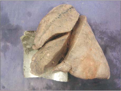
Fig. 1 Left lung with three lobes and two fissures.
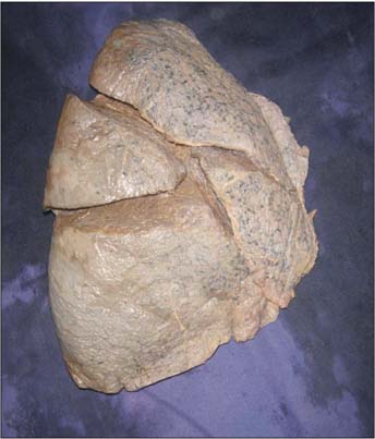
Fig. 2 Right lung with four lobes and three fissures.
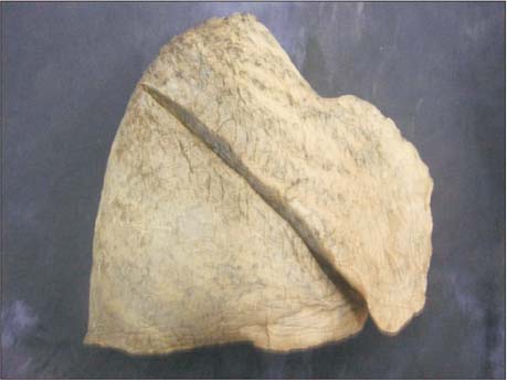
Fig. 3 Right lung with two lobes and one fissure.
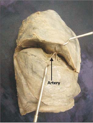
Fig. 4 Visible artery in the fissure.
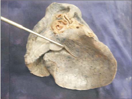
Fig. 5 Oblique fissure extending on to the base.
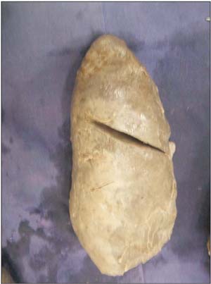
Fig. 6 Incomplete oblique fissure.
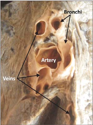
Fig. 7 Hilum of the right lung with three veins, one artery, and two bronchi.
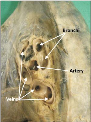
Fig. 8 Hilum of the right lung with four veins, two bronchi, and one artery.
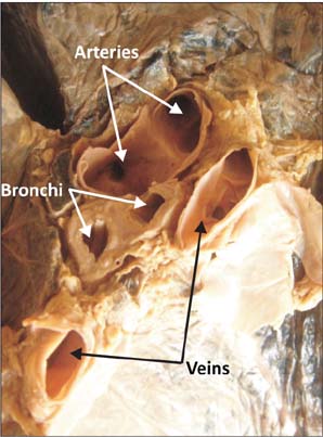
Fig. 9 Hilum of the left lung with two veins, two arteries, and two bronchi.
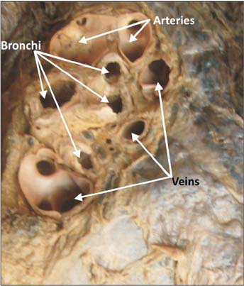
Fig. 10 Hilum of the left lung with three veins, two arteries, and four bronchi.
Reference
-
1. Shah P, Johnson D, Standring S. Thorax. In : Standring S, editor. Gray's Anatomy: The Anatomical Basis of Clinical Practice. 39th ed. Edinburgh: Churchill Livingstone;2005. p. 1068–1069.2. Rosse C, Gaddum-Rosse P. Hollinshead's textbook of anatomy. Philadelphia: Lipincott Williams & Wilkins;1997. p. 441–461.3. Brahmbhatt RJ, Chauhan KB, Bansal M, Brahmbhatt JN. Cadaveric study of azygous lobe of lung. Int J Basic Appl Med Sci. 2013; 3:30–33.4. Meenakshi S, Manjunath KY, Balasubramanyam V. Morphological variations of the lung fissures and lobes. Indian J Chest Dis Allied Sci. 2004; 46:179–182.5. Speckman JM, Gamsu G, Webb WR. Alterations in CT mediastinal anatomy produced by an azygos lobe. AJR Am J Roentgenol. 1981; 137:47–50.6. Aldur MM, Denk CC, Celik HH, Tasçioglu AB. An accessory fissure in the lower lobe of the right lung. Morphologie. 1997; 81:5–7.7. Tarver RD. How common are incomplete pulmonary fissures, and what is their clinical significance? AJR Am J Roentgenol. 1995; 164:761.8. Larsen WJ. Human embryology. New York: Churchill Livingstone;1993. p. 111–130.9. Polaczek M, Religioni J, Orłowski T. Anatomic variations of pulmonary vessels relevant with regard to lung tissue resections: literature review and personal experiences. Kardiochir Torakochirurgia Pol. 2013; 10:232–238.10. Prakash , Bhardwaj AK, Shashirekha M, Suma HY, Krishna GG, Singh G. Lung morphology: a cadaver study in Indian population. Ital J Anat Embryol. 2010; 115:235–240.11. Nene AR, Gajendra KS, Sarma MV. Lung lobes and fissures: a morphological study. Anatomy. 2011; 5:30–38.12. Ghosh E, Basu R, Dhur A, Roy A, Roy H, Biswas A. Variations of fissures and lobes in human lungs: a multicentric cadaveric study from West Bengal, India. Int J Anat Radiol Surg. 2013; 2:5–8.13. Sharma G, Vijayvergiya T. Anatomical variations in lobar pattern of the lungs: anatomical study and clinical significance. J Pharm Biomed Sci. 2013; 26:301–303.14. Mayuri J, Pradeep P, Vasudha N, Aparna T, Smita M. Anomalous lobar pattern of right lung: a case report. J Res Med Den Sci. 2013; 1:80–81.15. Berkmen T, Berkmen YM, Austin JH. Accessory fissures of the upper lobe of the left lung: CT and plain film appearance. AJR Am J Roentgenol. 1994; 162:1287–1293.16. Frija J, Schmit P, Katz M, Vadrot D, Laval-Jeantet M. Computed tomography of the pulmonary fissures: normal anatomy. J Comput Assist Tomogr. 1982; 6:1069–1074.17. Medlar EM. Variations in interlobar fissures. Am J Roentgenol Radium Ther. 1947; 57:723–725.18. Otsuji H, Uchida H, Maeda M, Iwasaki S, Yoshiya K, Hatakeyama M, Ohishi H, Iioka S, Kitamura S, Narita N. Incomplete interlobar fissures: bronchovascular analysis with CT. Radiology. 1993; 187:541–546.19. Abe S, Yamamoto M, Noguchi T, Yoshimoto T, Kinoshita H, Matsunaga S, Murakami G, Rodríguez-Vázquez JF. Fetal development of the minor lung segment. Anat Cell Biol. 2014; 47:12–17.20. Speckman JM, Gamsu G, Webb WR. Alterations in CT mediastinal anatomy produced by an azygos lobe. AJR Am J Roentgenol. 1981; 137:47–50.21. Aldur MM, Denk CC, Celik HH, Tasçioglu AB. An accessory fissure in the lower lobe of the right lung. Morphologie. 1997; 81:5–7.22. Esomonu UG, Taura MG, Modibbo MH, Egwu AO. Variation in the lobar pattern of the right and left lungs: A case report. Australas Med J. 2013; 6:511–514.
- Full Text Links
-
- Actions
-
Cited
- CITED
-
- Close
- Share
-
- Similar articles
-
- Variations in human pulmonary fissures and lobes: a study conducted in nepalese cadavers
- Anatomical variations and developmental anomalies of the thyroid gland in Ethiopian population: a cadaveric study
- Study on branching pattern of aortic arch in Indian
- Variations of azygos vein: a cadaveric study with clinical relevance
- Variations of the Occipital Sinus in Korean Adults
