J Periodontal Implant Sci.
2013 Feb;43(1):41-46. 10.5051/jpis.2013.43.1.41.
Effect of microthreads on removal torque and bone-to-implant contact: an experimental study in miniature pigs
- Affiliations
-
- 1Seoul National University School of Dentistry, Seoul, Korea.
- 2Department of Periodontology, Dental Research Institute, Seoul National University School of Dentistry, Seoul, Korea. periokoo@snu.ac.kr
- 3Implant R&D Center, Osstem Implant Co., Busan, Korea.
- KMID: 1783677
- DOI: http://doi.org/10.5051/jpis.2013.43.1.41
Abstract
- PURPOSE
The objective of this study was to evaluate the effect of microthreads on removal torque and bone-to-implant contact (BIC).
METHODS
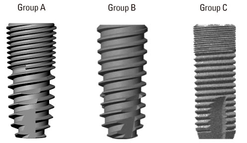
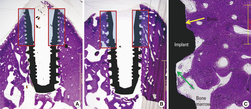
Twelve miniature pigs for each experiment, a total of 24 animals, were used. In the removal torque analysis, each animal received 2 types of implants in each tibia, which were treated with sandblasting and acid etching but with or without microthreads at the marginal portion. The animals were sacrificed after 4, 8, or 12 weeks of healing. Each subgroup consisted of 4 animals, and the tibias were extracted and removal torque was measured. In the BIC analysis, each animal received 3 types of implants. Two types of implants were used for the removal torque test and another type of implant served as the control. The BIC experiment was conducted in the mandible of the animals. The P1-M1 teeth were extracted, and after a 4-month healing period, 3 each of the 2 types of implants were placed, with one type on each side of the mandible, for a total of 6 implants per animal. The animals were sacrificed after a 2-, 4-, or 8-week healing period. Each subgroup consisted of 4 animals. The mandibles were extracted, specimens were processed, and BIC was analyzed.
RESULTS
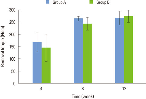
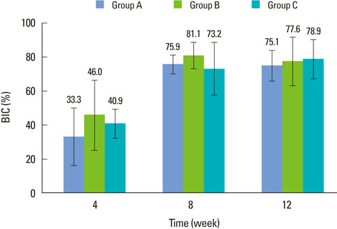
No significant difference in removal torque value or BIC was found between implants with and without microthreads. The removal torque value increased between 4 and 8 weeks of healing for both types of implants, but there was no significant difference between 8 and 12 weeks. The percentage of BIC increased between 2 and 4 weeks for all types of implants, but there was no significant difference between 4 and 8 weeks.
CONCLUSIONS
The existence of microthreads was not a significant factor in mechanical and histological stability.
Keyword
Figure
Reference
-
1. Klokkevold PR, Nishimura RD, Adachi M, Caputo A. Osseointegration enhanced by chemical etching of the titanium surface: a torque removal study in the rabbit. Clin Oral Implants Res. 1997. 8:442–447.
Article2. Carlsson L, Rostlund T, Albrektsson B, Albrektsson T. Removal torques for polished and rough titanium implants. Int J Oral Maxillofac Implants. 1988. 3:21–24.3. Shalabi MM, Wolke JG, Jansen JA. The effects of implant surface roughness and surgical technique on implant fixation in an in vitro model. Clin Oral Implants Res. 2006. 17:172–178.
Article4. Lee DW, Choi YS, Park KH, Kim CS, Moon IS. Effect of microthread on the maintenance of marginal bone level: a 3-year prospective study. Clin Oral Implants Res. 2007. 18:465–470.
Article5. Abrahamsson I, Berglundh T. Tissue characteristics at microthreaded implants: an experimental study in dogs. Clin Implant Dent Relat Res. 2006. 8:107–113.
Article6. Steigenga JT, al-Shammari KF, Nociti FH, Misch CE, Wang HL. Dental implant design and its relationship to long-term implant success. Implant Dent. 2003. 12:306–317.
Article7. Hudieb MI, Wakabayashi N, Kasugai S. Magnitude and direction of mechanical stress at the osseointegrated interface of the microthread implant. J Periodontol. 2011. 82:1061–1070.
Article8. Rasmusson L, Kahnberg KE, Tan A. Effects of implant design and surface on bone regeneration and implant stability: an experimental study in the dog mandible. Clin Implant Dent Relat Res. 2001. 3:2–8.
Article9. Misch CE. Contemporary implant dentistry. 2008. 3rd ed. St. Louis: Mosby Elsevier.10. Chung SH, Heo SJ, Koak JY, Kim SK, Lee JB, Han JS, et al. Effects of implant geometry and surface treatment on osseointegration after functional loading: a dog study. J Oral Rehabil. 2008. 35:229–236.
Article11. Ma P, Liu HC, Li DH, Lin S, Shi Z, Peng QJ. Influence of helix angle and density on primary stability of immediately loaded dental implants: three-dimensional finite element analysis. Zhonghua Kou Qiang Yi Xue Za Zhi. 2007. 42:618–621.12. Chun HJ, Cheong SY, Han JH, Heo SJ, Chung JP, Rhyu IC, et al. Evaluation of design parameters of osseointegrated dental implants using finite element analysis. J Oral Rehabil. 2002. 29:565–574.
Article13. Kong L, Liu BL, Hu KJ, Li DH, Song YL, Ma P, et al. Optimized thread pitch design and stress analysis of the cylinder screwed dental implant. Hua Xi Kou Qiang Yi Xue Za Zhi. 2006. 24:509–512. 515
- Full Text Links
- Actions
-
Cited
- CITED
-
- Close
- Share
- Similar articles
-
- The Biological Effects of Calcium Phosphate Coated Implant for Osseointegration in Beagle Dogs
- The influence of systemically administered oxytocin on the implant-bone interface area: an experimental study in the rabbit
- Histomorphometric and Removal Torque Values Comparision of Rough Surface Titanium Implants
- The influence of intentional mobilization of implant fixtures before osseointegration
- Influence of implant diameter on the osseointegration of implants; an experimental study in rabbits


