Extrahepatic Biliary Schwannomas: A Case Report
- Affiliations
-
- 1Digestive Disease Center, Kyunghee East-West Neo Medical Center, 149 Sangil-dong, Gangdong-gu, Seoul, Korea. krjoo@khu.ac.kr
- 2Department of Pathology, Kyung Hee University College of Medicine, Seoul, Korea.
- 3Department of Surgery, Kyung Hee University College of Medicine, Seoul, Korea.
- KMID: 1778369
- DOI: http://doi.org/10.3346/jkms.2007.22.3.549
Abstract
- Benign schwannomas arise in neural crest-derived Schwann cells. They can occur almost anywhere in the body, but their most common locations are the central nervous system, extremities, neck, mediastinum, and retroperitoneum. Schwannomas occurring in the biliary tract are extremely rare and mostly present with obstructive jaundice. We recently experienced a case of extrahepatic biliary schwannomas in a 64-yr-old female patient who presented with intra- and extrahepatic bile duct and gallbladder stones during a screening program. To the best of our knowledge, extrahepatic biliary schwannomas associated with bile duct stones have not been reported previously in the literature.
MeSH Terms
Figure
-
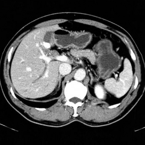
Fig. 1 Computed tomographic scan shows mildly dilated intrahepatic bile duct and many small stones in the gallbladder.
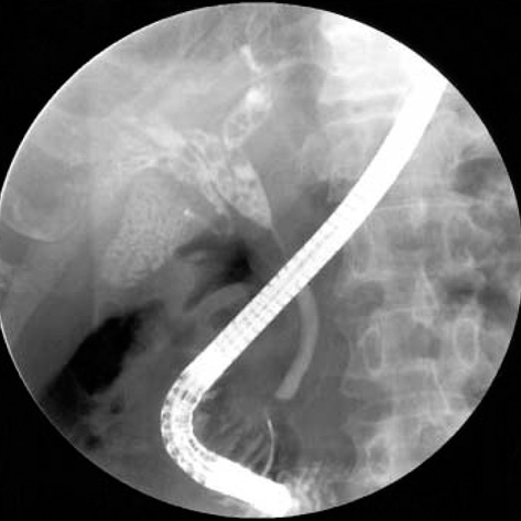
Fig. 2 Endoscopic retrograde cholangiography shows a focal stricture of the proximal common bile duct and an upstream bile duct dilatation with stones. The cystic duct is inserted aberrantly into the right intrahepatic duct. Gallbladder stones are also observed.
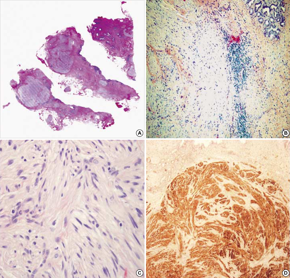
Fig. 3 (A) The specimen shows several well-demarcated nodules in the bile duct wall. (B) A nodule shows a typical pattern of a schwannoma consisting of spindle cells. Lymphocyte cuffing is also seen in the margin of the tumor (H&E ×100). (C) Magnified view shows interlacing fascicles with nuclear palisading (H&E ×200). (D) Immunochemical staining is positive for the S-100 protein (×100).
Reference
-
1. Malka D, Boige V, Dromain C, Debaere T, Pocard M, Ducreux M. Biliary tract neoplasm: update 2003. Curr Opin Oncol. 2004. 16:364–371.2. Northover JM, Terblanche J. A new look at the arterial supply of the bile duct in man and its surgical implications. Br J Surg. 1979. 66:379–384.
Article3. Enzinger FM, Weiss SW. Soft tissue tumors. 1983. 1nd ed. St Louis, Mo: Mosby-Year Book;586–597.4. Sarlomo-Rikala M, Miettinen M. Gastric schwannoma: a clinicopathological analysis of six cases. Histopathology. 1995. 27:355–360.5. Miettinen M, Shekitka KM, Sobin LH. Schwannomas in the colon and rectum: a clinicopathologic and immunohistochemical study of 20 cases. Am J Surg Pathol. 2001. 25:846–855.6. Honjo Y, Kobayashi Y, Nakamura T, Kakehira Y, Kitagqwa M, Ikematsu Y, Ozawa T, Nakamura H. Extrahepatic biliary schwannoma. Dig Dis Sci. 2003. 48:2221–2226.7. Oden B. Neurinoma of the common bile duct: report of a case. Acta Chir Scand. 1955. 108:393–397.8. Nagafuchi Y, Mitsuo H, Takeda S, Ohsato K, Tsuneyoshi M, Enjoji M. Benign schwannoma in the hepatoduodenal ligament: report of a case. Surg Today. 1993. 23:68–72.
Article9. Otani T, Shioiri T, Mishima H, Ishihara A, Maeshiro T, Matsuo A, Umekita N, Warabi M. Bile duct schwannoma developed in the remnant choledochal cyst-a case associated with total agenesis of the dorsal pancreas. Dig Liver Dis. 2005. 37:705–708.
Article10. Jakobs R, Albert J, Schilling D, Nuesse T, Riemann JF. Schwannoma of the common bile duct: a rare cause of obstructive jaundice. Endoscopy. 2003. 35:695–697.
Article11. Shiesh SC, Chen CY, Lin XZ, Liu ZA, Tsao HC. Melatonin prevents pigment gallstone formation induced by bile duct ligation in guinea pigs. Hepatology. 2000. 32:455–460.
Article12. Daimaru Y, Kido H, Hashimoto H, Enjoji M. Benign schwannoma of the gastrointestinal tract: a clinicopathologic and immunohistochemical study. Hum Pathol. 1988. 19:257–264.
Article13. Lasota J, Wasag B, Dansonka-Mieszkowska A, Karcz D, Millward CL, Rys J, Stachura J, Sobin LH, Miettinen M. Evaluation of NF2 and NF1 tumor suppressor genes in distinctive gastrointestinal nerve sheath tumors traditionally diagnosed as benign schwannomas: study of 20 cases. Lab Invest. 2003. 83:1361–1371.14. Levy AD, Quiles AM, Miettinen M, Sobin LH. Gastrointestinal schwannoma: CT features with clinicopathological correlation. Am J Roentgenol. 2005. 184:797–802.15. Rha SE, Byun JY, Jung SE, Chun HJ, Lee HG, Lee JM. Neurogenic tumors in the abdomen: tumor types and imaging characteristics. Radiographics. 2003. 23:29–43.
Article
- Full Text Links
-
- Actions
-
Cited
- CITED
-
- Close
- Share
-
- Similar articles
-
- Extrahepatic Bile Duct Duplication with Intraductal Papillary Neoplasm: A Case Report
- Biliary Web: A Rare Cause of Extrahepatic Biliary Obstruction
- Extrahepatic Biliary Atresia
- Restrictive Cardiomyopathy in a Patient with Extrahepatic Biliary Atresia
- A Case of Congenital Agenesis of the Gallbladder without Biliary Atresia associated with Duodenal Web
