Risk Factors for Adjacent Segment Disease after Posterolateral Lumbar Fusion
- Affiliations
-
- 1Department of Orthopaedic Surgery, College of Medicine, Dong-A University, Busan, Korea. gylee@dau.ac.kr
- KMID: 1747201
- DOI: http://doi.org/10.4184/jkss.2008.15.3.174
Abstract
-
STUDY DESIGN: This is a retrospective study.
OBJECTIVE
We wanted to analyze the treatment outcome and the risk factors for adjacent segment disease after lumbar fusion. SUMMARY OF LITERATURE REVIEW: Biomechanical alterations likely play a primary role in causing adjacent segment disease. Radiographically apparent, asymptomatic adjacent segment disease is common after lumbar fusion, but this does not correlate with the functional outcomes.
MATERIALS AND METHODS
We reviewed 544 patients who underwent lumbar fusion at a minimum of 5-year follow-up between March 1993 and August 2006. Risk factors analysis was performed for 48 of 544 patients with adjacent segment disease and who were needed a second operation, and the treatment outcomes were assessed for 46 patients with a minimum 1-year follow-up after the second operation. The average interval to the second operation was 4.5 years, and the average follow-up after the second operation was 34.5 months. The treatment outcome was assessed by using the modified Brodsky criteria and the reoperation rate was assessed in relation to several risk factors.
RESULTS
Excellent and good operative results were obtained in 29 cases (63%) and bony fusion was achieved in 41 cases (89%). Of the risk factors we examined, multi-level fusion, a high grade of initial radiographic degeneration, the loss of physiologic lumbar lordosis and the involvement of degenerative scoliosis were associated with a high reoperation rate, with statistical significance. Age, gender, the initial diagnosis, the upper placement of the proximal screws and the extent to the sacrum were not correlated with the reoperation rate.
CONCLUSION
The treatment outcome was relatively satisfactory; however, the factors influencing the treatment outcome of the second operation still need to be considered. The fusion level, the initial radiographic degeneration, the preservation of lumbar lordosis and the involvement of degenerative scoliosis are considered to be risk factors for the failure of lumbar fusion.
MeSH Terms
Figure
-
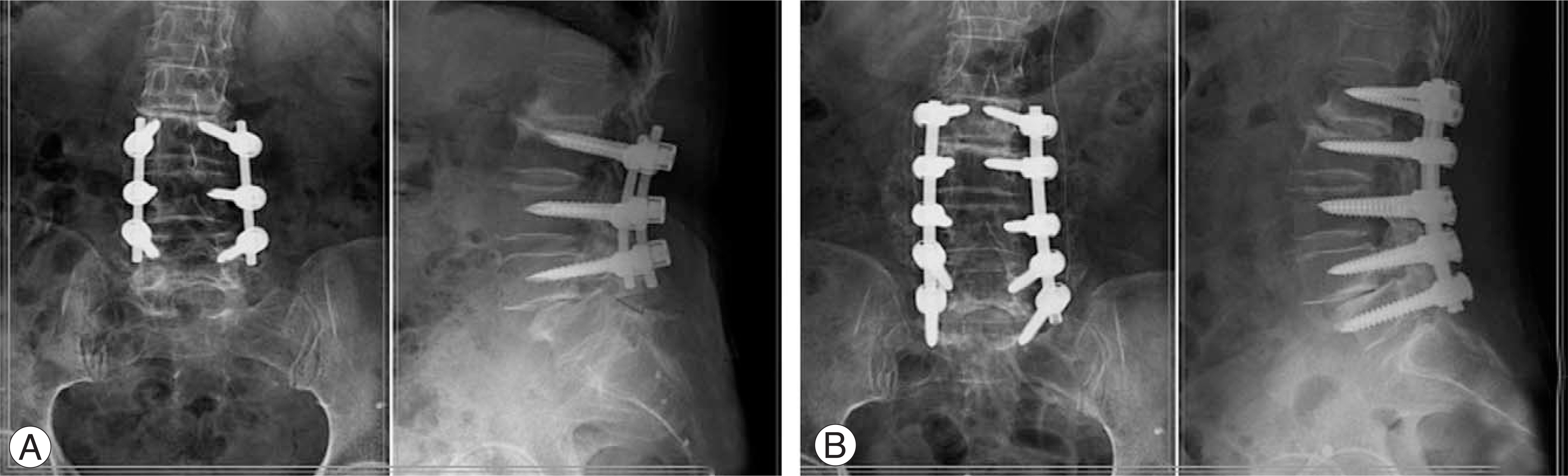
Fig. 1. Posterolateral fusion with instrumentation at L3-4-5 in a 62 year-old male patient. (A) Postoperative 2 years, plain radiographs show both upper and lower adjacent degenerative changes. (B) Extended posterior decompression and instrumented posterolateral fusion were performed. At the last follow-up, clinical result was good according to the modified Brodsky's criteria.
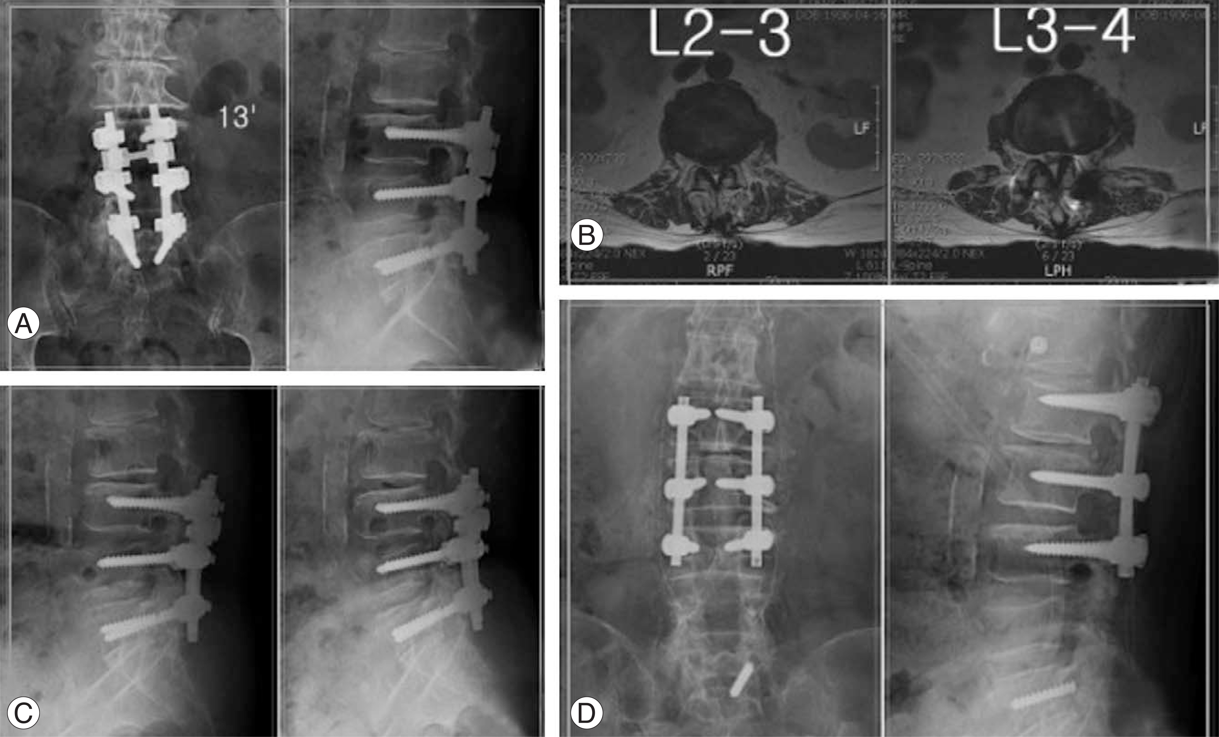
Fig. 2. Posterolateral fusion with instrumentation at L4-5-S1 in a 69 year-old female patient. (A) Postoperative 10 years, plain radiographs show degenerative lumbar scoliotic change about 13 degrees at AP view and adjacent degenerative changes at saggital view. S1 pedicle screw was broken. (B) T2-weight axial MR images show central and lateral recess type stenosis at L2-3 and L3-4. (C) Dynamic radiographs show L3-4 adjacent instability. (D) Metal removal and posterior decompression and instrumented posterolateral fusion were performed. At the last follow-up, clinical result was excellent according to the modified Brodsky's criteria.
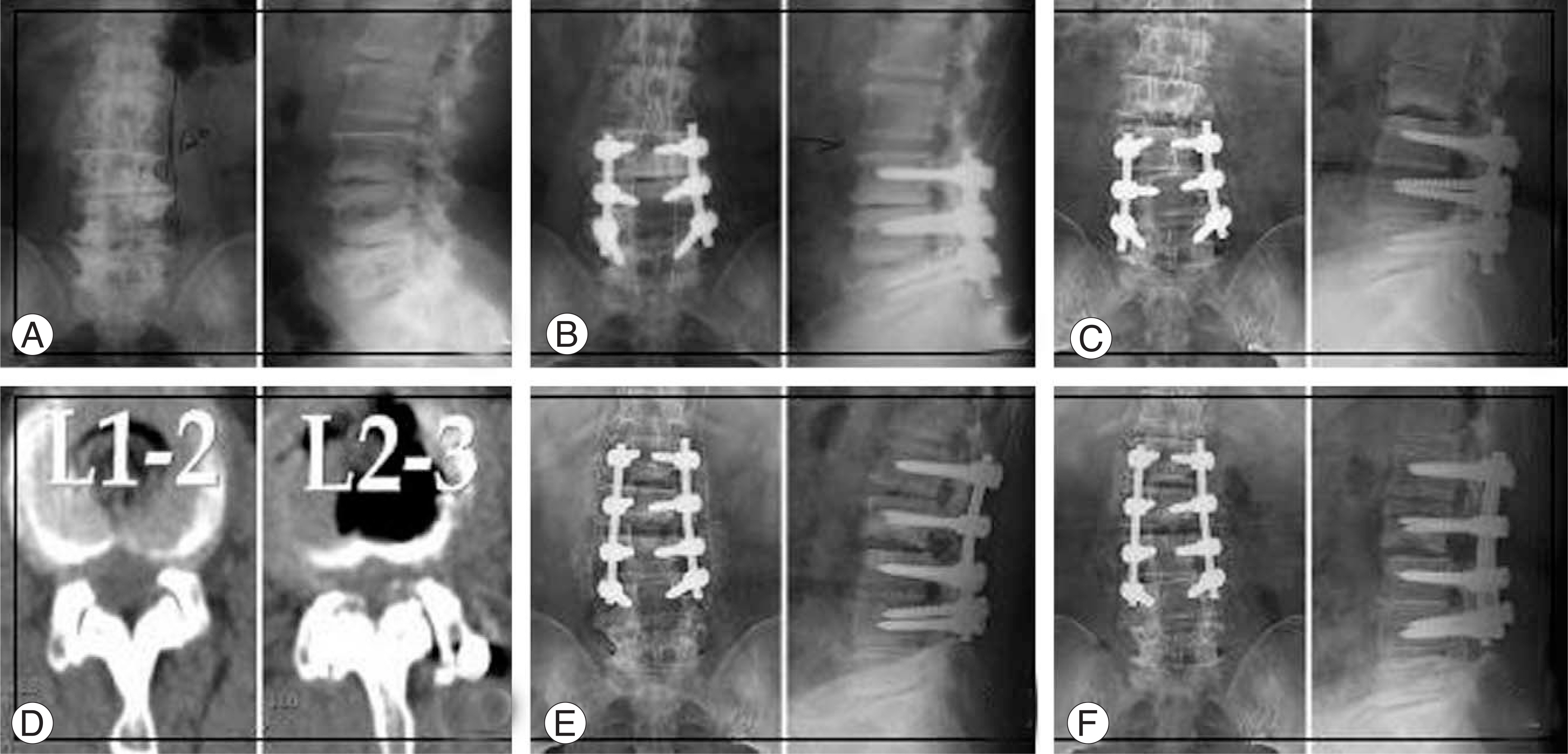
Fig. 3. A 64 year-old female radiographs. (A) Initial radiographs show multiple osteophytes and disc space narrowing with mild scoliotic change at L3-4-5. (B) Posterior decompression and instrumented posterolateral fusion were performed. Kellgren grade II degeneration and joint space widening were noted at L2-3 on immediate postoperative radiographs. (C) At postoperative 3 years, severe endplate degeneration, disc collapse and advanced scoliosis were noted at L2-3. (D) Computerized axial tomography images show central and lateral recess type stenosis at L1-2-3. (E) Extended posterior decompression and instrumented posterolateral fusion were performed. (F) At the last follow-up, radiographs show bone union and the clinical result was good according to the modified Brodsky's criteria.
Cited by 1 articles
-
Comparative Analysis of Revision Surgery Groups between within 5 Years and More than 10 Years after Lumbar Spinal Fusion Due to Adjacent Segment Disease
Jaewan Soh, Junghyeok Kim, Jae Chul Lee, Byung-Joon Shin
J Korean Orthop Assoc. 2016;51(3):214-220. doi: 10.4055/jkoa.2016.51.3.214.
Reference
-
1). Aota Y, Kumano K, Hirabayashi S. Postfusion instability at the adjacent segments after rigid pedicle screw fixation for degenerative lumbar spinal disorders. J Spinal Disord. 1995; 8:464–473.
Article2). Fritsch EW, Heisel J, Rupp S. The failed back surgery syndrome: reasons, intraoperative findings, and long-term results: a report of 182 operative treatments. Spine. 1996; 21:626–633.3). Frymoyer JW, Hanley E, Howe J, Kuhlmann D, Matteri R. A comparison of radiolographic findings in fusion and nonfusion patients ten or more years following lumbar disc surgery. Spine. 1979; 4:435–440.4). Guigui P, Lambert P, Lassale B, Deburge A. Long-term outcome at adjacent levels of lumbar arthrodesis. Rev Chir Orthop Reparatrice Appar Mot. 1997; 83:685–696.5). Ha KY, Sung TP. Changes of the adjacent mobile segment after cat spine fixation. J Kor Orthop Surg. 1997; 32:1808–1816.
Article6). Ha KY, Moon MS, Paek SY. Effect of Instumental stabilization and fusion of degenerative lumbar scoliosis on? unfused adjacent segment. J Kor Spine Surg. 1995; 2:270–278.7). Kumar MN, Jacquot F, Hall H. Long-term follow up of functional outcomes and radiographic changes at adjacent levels following lumbar spine fusion for degenerative disc disease. Eur Spine J. 2001; 10:309–313.8). Lehmann TR, Spratt KF, Tozzi JE, et al. Long-term follow-up of lower lumbar fusion patients. Spine. 1987; 12:97–104.
Article9). Rahm MD, Hall BB. Adjacent-segment degeneration after lumbar fusion with instrumentation: A retrospective study. J Spinal Disord. 1996; 9:392–400.10). Schlegel JD, Smith JA, Schleusener RL. Lumbar motion segment pathology adjacent to thoracolumbar, lumbar, and lumbosacral fusions. Spine. 1996; 21:970–981.
Article11). Etebar S, Cahill DW. Risk factors for adjacent-segment failure following lumbar fixation with rigid instrumentation for degenerative instability. J Neurosurg. 1999; 90:163–169.
Article12). Kim SS, Michelsen CB. Revision surgery for failed back surgery syndrome. Spine. 1992; 17:957–960.
Article13). Kumar MN, Baklanov A, Chopin D. Correlation between saggital plane changes and adjacent segment degeneration following lumbar spine fusion. Eur Spine J. 2001; 10:314–319.14). Lee CK, Langrana NA. Lumbosacral spinal fusion. A biomechanical study. Spine. 1984; 9:574–581.
Article15). Bastian L, Lange U, Knop C, Tusch G, Blauth M. Evaluation of the mobility of adjacent segments after posterior thoracolumbar fixation: A biomechanical study. Eur Spine J. 2001; 10:295–300.
Article16). Frymoyer JW, Hanley E, Howe J, Kuhlmann D, Matteri R. Disc excision and spine fusion in the management of lumbar disc disease. A minimum 10 year follow up. Spine. 1978; 13:1–6.17). Hambly MF, Wiltse LL, Raghavan N, Schneiderman G, Koenig C. The transition zone above a lumbosacral fusion. Spine. 1998; 23:1785–1792.
Article18). Whitecloud TS, Davis JM, Olive PM. Operative treatment of the degenerated segment adjacent to a lumbar fusion. Spine. 1994; 19:531–536.
Article19). Morgan FP, King T. Primary instability of lumbar vertebrae as a common cause of low back pain. J Bone Joint Surg. 1957; 39:6–22.
Article20). Dupuis PR, Yong-Hing K, Cassidy JD, Kirkaldy-Willis WH. Radiologic diagnosis of degenerative lumbar spinal instability. Spine. 1985; 10:262–276.
Article21). Ha KY, Schendel MJ, Lewis JL, Ogilvie JW. Effect of immobilization and configuration on lumbar adjacent segment biomechanics. J Spinal Disord. 1993; 6:99–105.22). Brunet JA, Wiley JJ. Acquired spondylolysis after spinal fusion. J Bone and Joint Surg. 1984; 66:720–724.
Article23). Ha KY, Kim KY, Park SJ, Lee YH. Changes of the adjacent-unfused mobile segment after instrumental lumbar fusion. More than 5 years follow-up. J Kor Spine Surg. 1998; 5:205–214.24). Chen WJ, Lai PL, Niu CC, Chen LH, Fu TS, Wong CB. Surgical treatment of adjacent instability after lumbar spine fusion. Spine. 2001; 26:519–524.
Article25). Hue KY, Zucherman J, White A, et al. Deterioration of motion segments adjacent to lumbar fusions. Annual meeting of North American Spine Society, Colorado Springs, Colorado. 1998.26). Shahin E, David W. Risk factors for adjacent-segment failure following lumbar fixation with rigid instrumentation for degenerative instability. J Neurosurg. 1999; 90:163–169.27). Kettler A, Wilke HJ, Haid C, Claes L. Effects of specimen length on the monosegmental motion behaviour of the lumbar spine. Spine. 2000; 25:543–550.28). Rohlman A, Neller S, Bergmann G, Graichen F, Claes L, Wilke HJ. Effect of an internal fixator and a bone graft on intersegmental spinal motion and intradiscal pressure in the adjacent regions. Eur Spine J. 2001; 10:301–308.
Article29). Cho JL, Park YS, Han JH, Lee CH, Roh WI. The changes of adjacent segments after spinal fusion. J Korean Spine Surg. 1998; 5:239–246.30). Ha KY, Kim YH, Kang KS. Surgery for adjacent segment changes after lumbosacral fusion. J Kor Spine Surg. 2002; 9:332–340.
Article31). Gillet P. The fate of the adjacent motion segment after lumbar fusion. J Spinal Disord Tech. 2003; 16:338–345.
- Full Text Links
-
- Actions
-
Cited
- CITED
-
- Close
- Share
-
- Similar articles
-
- Change of Segmental Motion After Lumbar Posterolateral Fusion
- Relation Between Adjacent Superior Segmental Disease and Facet Joint Violations After Posterolateral Lumbar Fusion
- Segmental Instability in Posterolateral Lumbar Spinal Fusion
- Adjacent Segment Pathology after Lumbar Spinal Fusion
- Degenerative Change of Adjacent Segments according to the Fusion Method after L4-5 Segmental Fusion: Comparative Study of Posterolateral Fusion and Posterior Lumbar Interbody Fusion
