Small Bowel Obstruction Caused by Peritoneal Immunoglobulin G4-Related Disease Mimicking Carcinomatosis: Case Report
- Affiliations
-
- 1Department of Diagnostic Radiology, Clinique St. Luc, Bouge, Namur 5004, Belgium. bcoulier@skynet.be
- 2Department of Internal Medicine, Clinique St. Luc, Bouge, Namur 5004, Belgium.
- 3Institute of Pathology and Genetics, Gosselies 6-6041, Belgium.
- 4Department of Visceral Surgery, Clinique St. Luc, Bouge, Namur 5004, Belgium.
- KMID: 1711479
- DOI: http://doi.org/10.3348/kjr.2014.15.1.66
Abstract
- We hereby report a case of diffuse pelvic peritoneal involvement by immunoglobulin G4-related disease (IgG4-RD). Numerous pelvic masses and nodules showing delayed enhancement on enhanced abdominal CT were found to congregate in the pelvic organs of a 57-year-old female presenting with intestinal subocclusion. The differentiation between peritoneal IgG4-RD and pelvic peritoneal carcinomatosis was only made by histopathology and immunohistochemistry performed after surgical resection. Autoimmune pancreatitis represents the historical prototype of IgG4-RD, but the spectrum of manifestations involving various organs has expanded during the last decade. In this report, we shortly review this clinical entity.
MeSH Terms
Figure
-
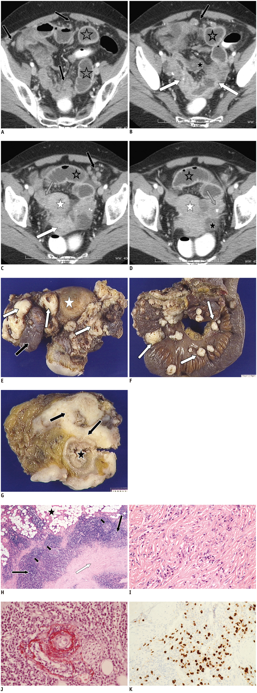
Fig. 1 57-year-old woman presenting with IgG4-related peritoneal disease. A-D.Four selected contrast-enhanced axial CT views of hypogastric and pelvic areas show diffuse infiltration of pelvic organs by enhancing masses which encase distal ileum (white arrows) causing intestinal subocclusion (grey stars). Diffuse small round nodules are also dispersed within peritoneal spaces (black arrows). White stars show uterus, black stars small amount of fluid in Douglas pouch and grey arrows ovaries. IgG4 = immunoglobulin G4 E. Gross anatomy of resected organs including uterus (white star), ovaries and portion of incarcerated ileum (black arrow). Organs are massively infiltrated by numerous irregular whitish nodules mimicking diffuse carcinomatosis (white arrows). F. Gross anatomy of 30 cm of resected ileum terminale. Numerous white nodules (white arrows) infiltrating mesenteric fat tissue. G. Section through ileum (black star) showing pseudotumoral fibrosing tissue (black arrows) infiltrating full thickness of intestinal wall. IgG4 = immunoglobulin G4 H. Photomicrograph (hematoxylin-eosin [H-E] stain; magnification, × 10) of section of fatty peritoneal tissue showing area of dense storiform fibrosis (white arrow) separated by dense inflammatory lymphoplasmacytic infiltrate (black arrows) with germinal center formation (small black arrows). Black star shows normal fat tissue. I. Photomicrographic (H-E stain; magnification, × 40) details of typical storiform fibrosis intermingled with lymphoplasmacytic inflammatory infiltrate. J. Photomicrographic (orcein stain; magnification, × 40) details of obliterative phlebitis. K. Photomicrograph (IgG4 immunostain; magnification, × 40) showing numerous IgG4-positive plasma cells. IgG4 = immunoglobulin G4
Reference
-
1. Divatia M, Kim SA, Ro JY. IgG4-related sclerosing disease, an emerging entity: a review of a multi-system disease. Yonsei Med J. 2012; 53:15–34.2. Ryu JH, Horie R, Sekiguchi H, Peikert T, Yi ES. Spectrum of Disorders Associated with Elevated Serum IgG4 Levels Encountered in Clinical Practice. Int J Rheumatol. 2012; 2012:232960.3. Hamano H, Kawa S, Horiuchi A, Unno H, Furuya N, Akamatsu T, et al. High serum IgG4 concentrations in patients with sclerosing pancreatitis. N Engl J Med. 2001; 344:732–738.4. Kim JH, Byun JH, Lee SS, Kim HJ, Lee MG. Atypical manifestations of IgG4-related sclerosing disease in the abdomen: imaging findings and pathologic correlations. AJR Am J Roentgenol. 2013; 200:102–112.5. Kamisawa T, Funata N, Hayashi Y, Eishi Y, Koike M, Tsuruta K, et al. A new clinicopathological entity of IgG4-related autoimmune disease. J Gastroenterol. 2003; 38:982–984.6. Vlachou PA, Khalili K, Jang HJ, Fischer S, Hirschfield GM, Kim TK. IgG4-related sclerosing disease: autoimmune pancreatitis and extrapancreatic manifestations. Radiographics. 2011; 31:1379–1402.7. Moh IH, Kim JB, Shin SR, Jung SW, Park SH, Kim JW, et al. A case of intraperitoneal immunoglobulin G4-related inflammatory pseudotumor. Korean J Gastroenterol. 2012; 60:258–261.8. Umehara H, Okazaki K, Masaki Y, Kawano M, Yamamoto M, Saeki T, et al. A novel clinical entity, IgG4-related disease (IgG4RD): general concept and details. Mod Rheumatol. 2012; 22:1–14.9. Stone JH, Khosroshahi A, Deshpande V, Chan JK, Heathcote JG, Aalberse R, et al. Recommendations for the nomenclature of IgG4-related disease and its individual organ system manifestations. Arthritis Rheum. 2012; 64:3061–3067.10. Yamashita H, Takahashi Y, Ishiura H, Kano T, Kaneko H, Mimori A. Hypertrophic pachymeningitis and tracheobronchial stenosis in IgG4-related disease: case presentation and literature review. Intern Med. 2012; 51:935–941.11. Inoue D, Zen Y, Abo H, Gabata T, Demachi H, Yoshikawa J, et al. Immunoglobulin G4-related periaortitis and periarteritis: CT findings in 17 patients. Radiology. 2011; 261:625–633.12. Choi JW, Kim SY, Moon KC, Cho JY, Kim SH. Immunoglobulin G4-related sclerosing disease involving the urethra: case report. Korean J Radiol. 2012; 13:803–807.13. Chen TS, Montgomery EA. Are tumefactive lesions classified as sclerosing mesenteritis a subset of IgG4-related sclerosing disorders? J Clin Pathol. 2008; 61:1093–1097.14. Minato H, Shimizu J, Arano Y, Saito K, Masunaga T, Sakashita T, et al. IgG4-related sclerosing mesenteritis: a rare mesenteric disease of unknown etiology. Pathol Int. 2012; 62:281–286.
- Full Text Links
-
- Actions
-
Cited
- CITED
-
- Close
- Share
-
- Similar articles
-
- Immunoglobulin G4-Related Lung Disease Mimicking Lung Cancer: Two Case Reports
- Erdheim–Chester Disease Involving the Biliary System and Mimicking Immunoglobulin G4-Related Disease: A Case Report
- A Case of Recurrent Ischemic Stroke Associated with Immunoglobulin G4-Related Disease
- A Case of Immunoglobulin G4-Related Sclerosing Disease Mimicking Lung Cancer
- A Case of Small Bowel Obstruction due to Peritoneal Encapsulation
