Adult Pilomyxoid Astrocytoma Mimicking a Cortical Brain Tumor: MR Imaging Findings
- Affiliations
-
- 1Department of Radiology, University of Ulsan College of Medicine, Ulsan University Hospital, Korea. ycweon@hanmail.net
- 2Department of Pathology, University of Ulsan College of Medicine, Ulsan University Hospital, Korea.
- 3Department of Neurosurgery, University of Ulsan College of Medicine, Ulsan University Hospital, Korea.
- KMID: 1460064
- DOI: http://doi.org/10.3348/jksr.2010.62.4.335
Abstract
- A pilomyxoid astrocytoma (PMA) is a recently identified low-grade neoplasm that was previously classified as a pilocytic astrocytoma (PA), yet demonstrates unique histological features and more aggressive behavior. Although a PMA is generally a tumor of early childhood and typically occurs in the hypothalamic/chiasmatic region, it can mimic cortical tumors, especially in adults. We report the MR findings of a PMA presenting as a cortical brain tumor in an adult with neurofibromatosis 1 (NF1).
MeSH Terms
Figure
-
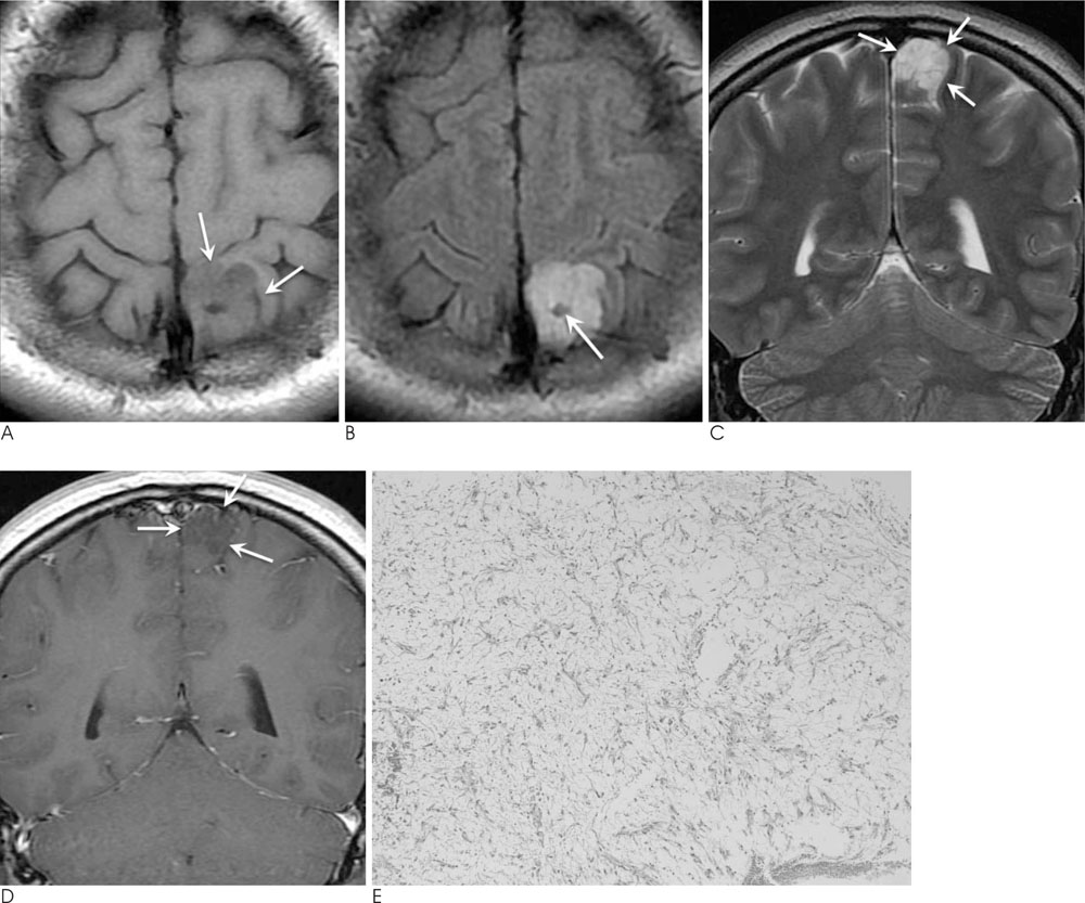
Fig. 1 A pilomyxoid astrocytoma in a 22-year-old woman presenting with a seizure. A. An axial, T1-weighted MR image shows a well-circumscribed, low-signal intensity solid mass in the cortex of the left parietal lobe (arrows). B. On FLAIR images, the mass is shown as high-signal intensity without hemorrhage or peritumoral edema. The cyst-like portion at the center of the mass is caused by the partial volume of the overlying sulcus of the parietal lobe (arrow). C. The mass appears to be located mainly in the cortex of the parietal lobe on the coronal, T2-weighted MR images (arrows). D. After gadolinium administration, there is no significant enhancement within the mass (arrows). E. A photomicrograph (original magnification, ×100; hematoxylin-eosin stain) shows that the tumor is composed of a monomorphic population of cells in a myxoid background. The tumor cells have round to oval nuclei and a spindle cytoplasmic process. There are no Rosenthal fibers or eosinophilic granular bodies. Also, no mitotic figure is seen.
Reference
-
1. Brat DJ, Parisi JE, Kleinschmidt-DeMasters BK, Yachnis AT, Montine TJ, Boyer PJ, et al. Surgical neuropathology update: a review of changes introduced by the WHO classification of tumours of the central nervous system, 4th edition. Arch Pathol Lab Med. 2008; 132:993–1007.2. Tihan T, Fisher PG, Kepner JL, Godfraind C, McComb RD, Goldthwaite PT, et al. Pediatric astrocytomas with monomorphous pilomyxoid features and a less favorable outcome. J Neuropathol Exp Neurol. 1999; 58:1061–1068.3. Komotar RJ, Burger PC, Carson BS, Brem H, Olivi A, Goldthwaite PT, et al. Pilocytic and pilomyxoid hypothalamic/chiasmatic astrocytomas. Neurosurgery. 2004; 54:72–79.4. Linscott LL, Osborn AG, Blaser S, Castillo M, Hewlett RH, Wieselthaler N, et al. Pilomyxoid Astrocytoma: expanding the Imaging Spectrum. AJNR Am J Neuroradiol. 2008; 29:1861–1866.5. Arslanoglu A, Cirak B, Horska A, Okoh J, Tihan T, Aronson L, et al. MR imaging characteristics of pilomyxoid astrocytomas. AJNR Am J Neuroradiol. 2003; 24:1906–1908.6. Komotar RJ, Mocco J, Zacharia BE, Wilson DA, Kim PY, Canoll PD, et al. Astrocytoma with pilomyxoid features presenting in an adult. Neuropathology. 2006; 26:89–93.7. Gottfried ON, Fults DW, Townsend JJ, Couldwell WT. Spontaneous hemorrhage associated with a pilomyxoid astrocytoma: case report. J Neurosurg. 2003; 99:416–420.8. Listernick R, Charrow J, Greenwald M, Mets M. Natural history of optic pathway tumors in children with neurofibromatosis type 1: a longitudinal study. J Pediatr. 1994; 125:63–66.9. Rodriguez FJ, Perry A, Gutmann DH, O'Neill BP, Leonard J, Bryant S, et al. Gliomas in neurofibromatosis type 1: a clinicopathologic study of 100 patients. J Neuropathol Exp Neurol. 2008; 67:240–249.10. Khanani MF, Hawkins C, Shroff M, Dirks P, Capra M, Burger PC, et al. Pilomyxoid astrocytoma in a patient with neurofibromatosis. Pediatr Blood Cancer. 2006; 46:377–380.
- Full Text Links
-
- Actions
-
Cited
- CITED
-
- Close
- Share
-
- Similar articles
-
- Juvenile Pilomyxoid Astrocytoma in the Opticohypothalamus
- Intermediate Pilomyxoid Astrocytoma in the Cerebellum of a 5-Year-Old Boy
- A Case of Anaplastic Astrocytoma in Term Pregnancy
- Intracranial Gossypiboma Mimicking a Recurrent Low Grade Astrocytoma: Case Report
- MR Imaging Findings of Gliosarcoma: Report of Three Cases
