Primary carcinosarcoma of the gallbladder
- Affiliations
-
- 1Department of Surgery, Kangwon National University Hospital, Chuncheon, Korea. skhong@knuh.or.kr
- KMID: 1437761
- DOI: http://doi.org/10.4174/jkss.2012.82.1.54
Abstract
- Carcinosarcoma of gallbladder (CSGB) is a rare malignancy characterized by malignant epithelial and mesenchymal components. Its pathogenesis is unknown and most CSGBs are associated with poor survival because the disease normally presents at an advanced stage, and as a result, curative resection is uncommon. This report describes a case that underwent curative resection. A 77-year-old woman presented with right upper quadrant pain. The preoperative diagnosis was gallbladder (GB) cancer, and thus, curative radical cholecystectomy was performed. However, pathologic examination of the surgical specimen revealed that the tumor was composed of two histologic components of squamous cell carcinoma and spindle cell sarcoma, which was consistent with a diagnosis of carcinosarcoma. The tumor was found to extend to the perimuscular connective tissue and to have metastasized to one lymph node (LN). The prognosis of CSGB remains poor despite curative resection, and thus, the authors recommend that effort be made to improve surgical outcomes.
Keyword
MeSH Terms
Figure
-
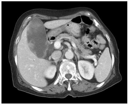
Fig. 1 Abdomen computed tomography showed diffuse distension of gallbladder (GB) with irregular intraluminal polypoid masses - possible GB cancer rather than xanthogranulomatous cholecystitis.
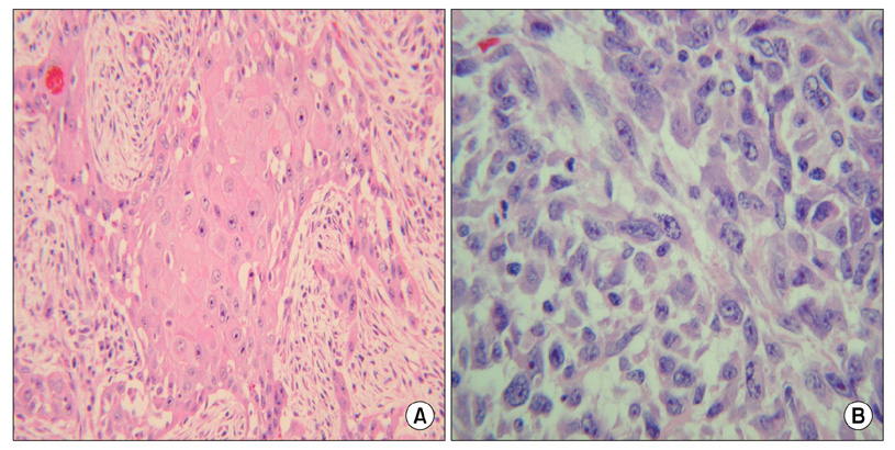
Fig. 2 Microscopic finding. (A) Well differentiated squamous cell carcinoma components (H&E, ×200). (B) High-grade spindle cell sarcoma components (H&E, ×400).
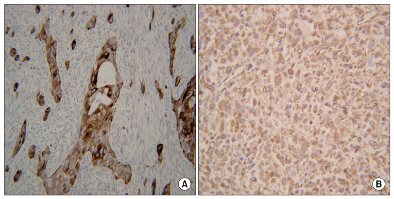
Fig. 3 Immunohistochemical stain. (A) Strong cytokeratin positivity in malignant glands forming the epithelial component (Cytokeratin, ×200). (B) Strong vimentin positivity in the sarcoma component (Vimentin, ×200).
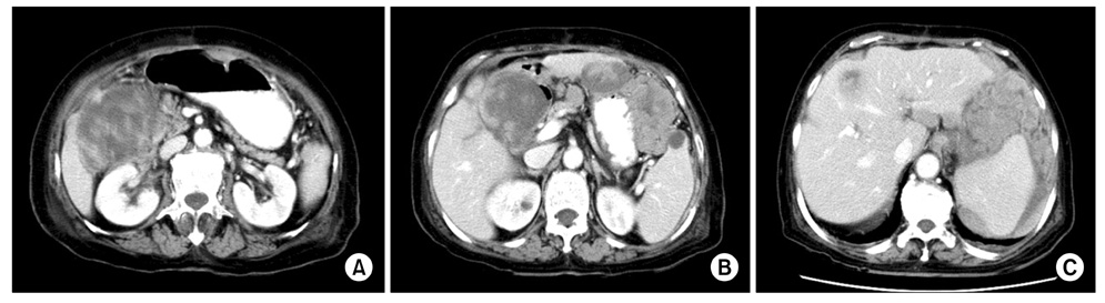
Fig. 4 Abdomen computed tomography showed huge metastatic mass involving liver S4 (A), and duodenum 1st portion (B). Multiple variable sized masses with central necrosis in the dependant portion of the abdominal cavity (C).
Reference
-
1. Baillie J. Tumors of the gallbladder and bile ducts. J Clin Gastroenterol. 1999. 29:14–21.2. Huguet KL, Hughes CB, Hewitt WR. Gallbladder carcinosarcoma: a case report and literature review. J Gastrointest Surg. 2005. 9:818–821.3. Okabayashi T, Sun ZL, Montgomey RA, Hanazaki K. Surgical outcome of carcinosarcoma of the gall bladder: a review. World J Gastroenterol. 2009. 15:4877–4882.4. Uzun MA, Koksal N, Gunerhan Y, Celik A, Guneş P. Carcinosarcoma of the gallbladder: report of a case. Surg Today. 2009. 39:168–171.5. Nishihara K, Tsuneyoshi M. Undifferentiated spindle cell carcinoma of the gallbladder: a clinicopathologic, immunohistochemical, and flow cytometric study of 11 cases. Hum Pathol. 1993. 24:1298–1305.6. Guo KJ, Yamaguchi K, Enjoji M. Undifferentiated carcinoma of the gallbladder. A clinicopathologic, histochemical, and immunohistochemical study of 21 patients with a poor prognosis. Cancer. 1988. 61:1872–1879.7. Liu KH, Yeh TS, Hwang TL, Jan YY, Chen MF. Surgical management of gallbladder sarcomatoid carcinoma. World J Gastroenterol. 2009. 15:1876–1879.8. Lumsden AB, Mitchell WE, Vohman MD. Carcinosarcoma of the gallbladder: a case report and review of the literature. Am Surg. 1988. 54:492–494.9. Hotta T, Tanimura H, Yokoyama S, Ura K, Yamaue H. So-called carcinosarcoma of the gallbladder; spindle cell carcinoma of the gallbladder: report of a case. Surg Today. 2002. 32:462–467.
