Pituitary Stalk Hemangioblastoma in a von Hippel-Lindau Patient : Clinical Course Follow-Up Over a 20-Year Period
- Affiliations
-
- 1Department of Radiology, College of Medicine, Kyung Hee University, Seoul, Korea.
- 2Department of Radiology, Kyung Hee University Hospital, Seoul, Korea. euijkim@hanmail.net
- 3Department of Neurosurgery, Kyung Hee University Hospital, Seoul, Korea.
- KMID: 1426198
- DOI: http://doi.org/10.3340/jkns.2013.53.5.297
Abstract
- Supratentorial hemangioblastomas (HBs) are rare, and pituitary stalk HBs are extremely uncommon; therefore, pituitary stalk evaluation is often overlooked. Herein, we report the development of pituitary stalk HB over a 20-year period and the importance of regular long-term follow up for patients with HBs.
Keyword
Figure
-
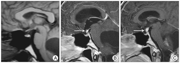
Fig. 1 Serial magnetic resonance imaging (MRI) of a pituitary stalk hemangioblastoma. A : Sagittal T1-weighted MRI performed 8 years following original resection reveals a normal pituitary stalk. B : Contrast-enhanced sagittal T1-weighted MRI performed 17 years following the initial surgery shows a 5-mm pituitary stalk hemangioblastoma (arrow). C : Twenty-two years following the initial surgery, MRI demonstrates an increased size of the pituitary stalk mass (arrow).
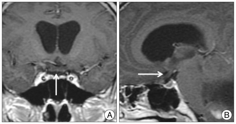
Fig. 2 Coronal (A) and sagittal (B) contrast-enhanced T1-weighted MRI reveals a thickened edematous optic chiasm (arrow).
Cited by 2 articles
-
Surgical Treatment of Hemangioblastoma in the Pituitary Stalk: An Extremely Rare Case
Jaejoon Lim, Sunghyun Noh, Kyung Gi Cho
Yonsei Med J. 2016;57(2):518-522. doi: 10.3349/ymj.2016.57.2.518.Sporadic Hemangioblastoma in the Pituitary Stalk: A Case Report and Review of the Literature
Gun-Ill Lee, Jae-Min Kim, Kyu-Sun Choi, Choong-Hyun Kim
J Korean Neurosurg Soc. 2015;57(6):465-468. doi: 10.3340/jkns.2015.57.6.465.
Reference
-
1. Kato M, Ohe N, Okumura A, Shinoda J, Nomura A, Shuin T, et al. Hemangioblastomatosis of the central nervous system without von Hippel-Lindau disease : a case report. J Neurooncol. 2005; 72:267–270. PMID: 15937651.
Article2. Kim HR, Suh YL, Kim JW, Lee JI. Disseminated hemangioblastomatosis of the central nervous system without von Hippel-Lindau disease : a case report. J Korean Med Sci. 2009; 24:755–759. PMID: 19654966.
Article3. Lonser RR, Butman JA, Kiringoda R, Song D, Oldfield EH. Pituitary stalk hemangioblastomas in von Hippel-Lindau disease. J Neurosurg. 2009; 110:350–353. PMID: 18834262.
Article4. Lonser RR, Glenn GM, Walther M, Chew EY, Libutti SK, Linehan WM, et al. von Hippel-Lindau disease. Lancet. 2003; 361:2059–2067. PMID: 12814730.
Article5. Peyre M, David P, Van Effenterre R, François P, Thys M, Emery E, et al. Natural history of supratentorial hemangioblastomas in von Hippel-Lindau disease. Neurosurgery. 2010; 67:577–587. discussion 587. PMID: 20647972.
Article
- Full Text Links
-
- Actions
-
Cited
- CITED
-
- Close
- Share
-
- Similar articles
-
- Sporadic Hemangioblastoma in the Pituitary Stalk: A Case Report and Review of the Literature
- Multifocal Spinal Hemangioblastoma in von Hippel-Lindau Syndrome: A Case Report and Literature Review
- Meningeal Supratentorial Hemangioblastoma in a Patient with Von Hippel-Lindau Disease Mimicking Angioblastic Menigioma
- Familial Occurrence of Von hippel-Lindau Disease: Case Report
- Brain Metastasis of Renal Cell Carcinoma in Von Hippel-Lindau Disease
