J Vet Sci.
2008 Dec;9(4):425-427. 10.4142/jvs.2008.9.4.425.
Eosinophilic myositis in a slaughtered Korean native cattle
- Affiliations
-
- 1Department of Veterinary Pathology, College of Veterinary Medicine, Kyungpook National University, Daegu 702-701, Korea. jeongks@knu.ac.kr
- 2College of Veterinary Medicine, Konkuk University, Seoul 143-701, Korea.
- KMID: 1104920
- DOI: http://doi.org/10.4142/jvs.2008.9.4.425
Abstract
- Histopathological findings of eosinophilic myositis in the carcass of a slaughtered Korean native cow are presented. Lesions contained massive fibrous septae with vacuolar changes in some lesions, and the hypercontraction and rupturing of muscle bundles, with replacement by eosinophils. Necrosis and severe eosinophil infiltration were observed. Sarcoplasmic fragmentation and atrophy developed. Typical of granuloma, calcified myofibers were focally surrounded by macrophages and numerous inflammatory cells, and multinucleated giant cell formation was evident.
Keyword
MeSH Terms
Figure
-
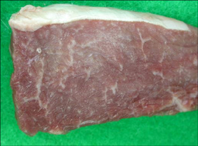
Fig. 1 Appearance of the skeletal muscle of the Korean native cow. The muscle was generally whitish and pale with a white streak.
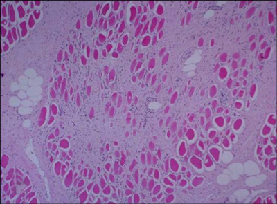
Fig. 2 Histochemical examination of the skeletal muscle of the Korean native cow. The whitish and pale muscle exhibited augmented interstitial fibrous septa and the strong activation of fibroblasts between the myofibers. H&E stain. ×12.
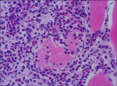
Fig. 3 Eosinophil examination of the skeletal muscle of the Korean native cow. Massive eosinophils are present within and adjacent to the affected myofibers. H&E stain. ×33.
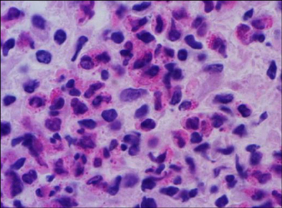
Fig. 4 High magnification of Fig. 3. ×66.
Reference
-
1. Bundza A, Feltmate TE. Eosinophilic myositis/lymphadenitis in slaughter cattle. Can Vet J. 1989. 30:514–516.2. Gajadhar AA, Yates WD, Allen JR. Association of eosinophilic myositis with an unusual species of Sarcocystis in a beef cow. Can J Vet Res. 1987. 51:373–378.3. Granstrom DE, Ridley RK, Baoan Y, Gershwin LJ. Immunodominant proteins of Sarcocystis cruzi bradyzoites isolated from cattle affected or nonaffected with eosinophilic myositis. Am J Vet Res. 1990. 51:1151–1155.4. Hall FC, Krausz T, Walport MJ. Idiopathic eosinophilic myositis. QJM. 1995. 88:581–586.5. Jensen R, Alexander AF, Dahlgren RR, Jolley WR, Marquardt WC, Flack DE, Bennett BW, Cox MF, Harris CW, Hoffmann GA, Troutman RS, Hoff RL, Jones RL, Collins JK, Hamar DW, Cravans RL. Eosinophilic myositis and muscular sarcocystosis in the carcasses of slaughtered cattle and lambs. Am J Vet Res. 1986. 47:587–593.6. Pickering MC, Walport MJ. Eosinophilic myopathic syndromes. Curr Opin Rheumatol. 1998. 10:504–510.
Article7. Reiten AC, Jensen R, Griner LA. Eosinophilic myositis (sarcosporidiosis; sarco) in beef cattle. Am J Vet Res. 1966. 27:903–906.8. Thomas JH. Jubb KVF, Kennedy PC, Palmer N, editors. Muscle and tendon. Pathology of Domestic Animals. 1985. Vol. 1:3rd ed. Orlando: Academic Press;245–252.9. Trüeb RM, Lubbe J, Torricelli R, Panizzon RG, Wüthrich B, Burg G. Eosinophilic myositis with eosinophilic cellulitislike skin lesions. Association with increased serum levels of eosinophil cationic protein and interleukin-5. Arch Dermatol. 1997. 133:203–206.
Article10. Wouda W, Snoep JJ, Dueby JP. Eosinophilic myositis due to Sarcocystis hominis in a beef cow. J Comp Pathol. 2006. 135:249–253.
Article
- Full Text Links
-
- Actions
-
Cited
- CITED
-
- Close
- Share
-
- Similar articles
-
- Epizootiological and Bacteriological Studies of Bovine Tuberculosis in Korea -(Report 4) A Study on the Atypical Mycobacteria Isolated from Native Korean Cattle-
- Focal eosinophilic myositis presenting with leg pain and tenderness
- Focal Eosinophilic Myositis
- Eosinophilic Myositis Induced by Anti-tuberculosis Medication
- Epitheliogenesis imperfecta in a bovine fetus of Korean native cattle
