The Poor Outcome of the Delayed Diagnosis of Acute Spontaneous Spinal Epidural Hematoma: Two Cases Report
- Affiliations
-
- 1Department of Orthopaedic Surgery, College of Medicine, Chonbuk National University Medical School, Chonju, Korea. osdr2815@chol.com
- 2Institute for Medical Science, Chonbuk National University Medical School, Chonju, Korea.
- KMID: 1095215
- DOI: http://doi.org/10.3346/jkms.2005.20.2.331
Abstract
- We present two patients who had acute paraplegia with sensory loss due to spontaneous spinal epidural hematoma (SSEH). One had myocardial infraction and the other had deep vein thrombosis, and the former was treated with anticoagulants and the latter was treated with thrombolytic agent. We analyzed the neurological status of our two cases each between its preoperative and postoperative state. Postoperatively both showed no improvement of neurologic symptom, and on follow-up of 12 months, one showed no neurologic improvement and the other showed a insignificant improvement of lower extremity muscle power (trace knee extensor/ankle dorsi-flexor). We thought that this poor outcome was due to delayed operation, which was done more than 24 hr after the symptom onset. The outcome in SSEH is essentially determined by the time taken from symptom onset to operation. Therefore, early and precise diagnosis such as careful history taking and MRI evaluation is necessary.
MeSH Terms
Figure
-
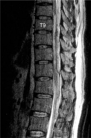
Fig. 1 Saggital T2 weighted MR Image shows the heterogenous hyperintense lesion with focal areas of hypointensity posterior to the compressed spinal cord in T8-L2 level.
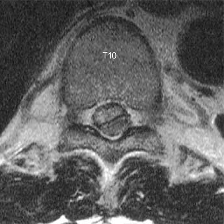
Fig. 2 Axial T2 weighted MR image shows the spinal cord compressed by the hyperintense lesion posterior to it in T10 level.
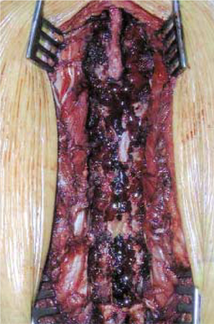
Fig. 3 Operative finding: Spinal cord is compressed by extensive blood clot and old hematoma. No other vascular abnormality.
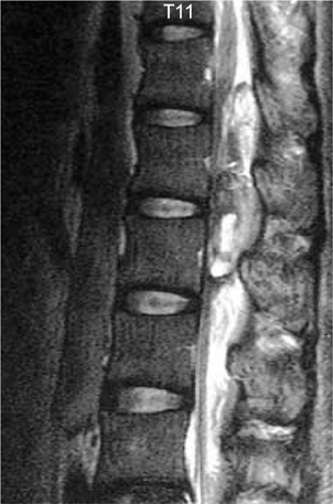
Fig. 4 Saggital T2 weighted MR Image shows the heterogenous hyperintense lesion with focal areas of hypointensity posterior to the compressed spinal cord in T12-L2 level.

Fig. 5 Axial T2 weighted MR image shows the spinal cord compressed by the hyperintense lesion posterior to it in T12 level.
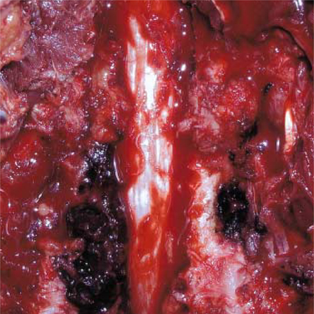
Fig. 6 Operative finding: The spinal cord is compressed by hematoma and blood clot. No other vascular abnormality.
Reference
-
1. Awada A, Russell N, Fayez NA, Naufal R, Kohlani HA. Spontaneous cervical epidural hematoma: case report. Spinal Cord. 1998. 36:71–72.
Article2. Hayem G, Deutsch E, Roux S, Palazzo E, Grossin M, Meyer O. Spontaneous spinal epidural hematoma with spinal cord compression complicating plasma cell myeloma: a case report. Spine. 1998. 23:2432–2435.3. Alexiadou-Rudolf C, Ernestus RI, Nanassis K, Lanfermann H, Klug N. Acute nontraumatic spinal epidural hematomas: An important differential diagnosis in spinal emergencies. Spine. 1998. 23:1810–1813.4. Chang H, Bahk WJ, Park SA. Traumatic epidural hematoma of the cervical spine. A case report. J Korean Spine Surg. 1996. 3:262–266.5. Lonjon MM, Paquis P, Chanalet S, Grellier P. Nontraumatic spinal epidural hematoma: report of four cases and review of the literature. Neurosurgery. 1997. 41:483–487.
Article6. Groen RJ, Van Alphen HA. Operative treatment of spontaneous spinal epidural hematomas: a study of the factors determining postoperative outcome. Neurosurgery. 1996. 39:494–509.
Article7. Lawton MT, Porter RW, Heiserman JE, Jacobowitz R, Sonntag VK, Dickman CA. Surgical management of spinal epidural hematoma: relationship between surgical timing and neurological outcome. J Neurosurg. 1995. 83:1–7.
Article
- Full Text Links
-
- Actions
-
Cited
- CITED
-
- Close
- Share
-
- Similar articles
-
- Spontaneous Lumbar Spinal Epidural Hematoma without Rist Factors: A Case Report
- Spontaneous Spinal Epidural Hematoma
- Surgical Treatment of Spontaneous Spinal Epidural Hematoma: A Case Report
- Spontaneous Spinal Epidural Hematoma Mimicking Lumbar Disc Herniation
- Acute and Delayed Epidural Hematoma After Total Spondylectomy for a Metastatic Spinal Tumor: A Case Report
