Gender Differences in Diagnostic Values of Visceral Fat Area and Waist Circumference for Predicting Metabolic Syndrome in Koreans
- Affiliations
-
- 1Divisions of Endocrinology and Metabolism, Department of Internal Medicine, Seoul National University College of Medicine, Seoul, Korea. ymchomd@snu.ac.kr
- 2Department of Molecular Medicine and Biopharmaceutical Sciences, Graduate School of Convergence Science and Technology, Seoul National University, Seoul, Korea.
- KMID: 1094262
- DOI: http://doi.org/10.3346/jkms.2011.26.7.906
Abstract
- Abdominal fat accumulation is known to be strongly implicated in development of metabolic syndrome (MetS). We examined diagnostic values of obesity-related parameters in 95 men and 185 women, and we determined optimal cutoff values of visceral fat area (VFA) and waist circumference (WC) for predicting the presence of multiple non-adipose components of MetS. Receiver operating characteristic (ROC) curve analysis revealed that VFA was the best indicator of MetS. WC and VFA exhibited similar diagnostic values for men and postmenopausal women, whereas WC was inferior to VFA for premenopausal women (area under ROC curve of VFA and WC was 0.76 and 0.52, respectively; P < 0.001). Optimal cutoff points of VFA and WC for predicting MetS were 136 cm2 and 89 cm in men and 95 cm2 and 82 cm in women, respectively. Subjects with VFA and WC above these cutoff values exhibited increased insulin resistance and increased carotid intima-media thickness. In conclusion, WC has a diagnostic value similar to VFA for predicting MetS in men and postmenopausal women, but not in premenopausal women. Further studies are necessary to develop a simple clinical parameter that reflects visceral fat in premenopausal women.
MeSH Terms
Figure
-
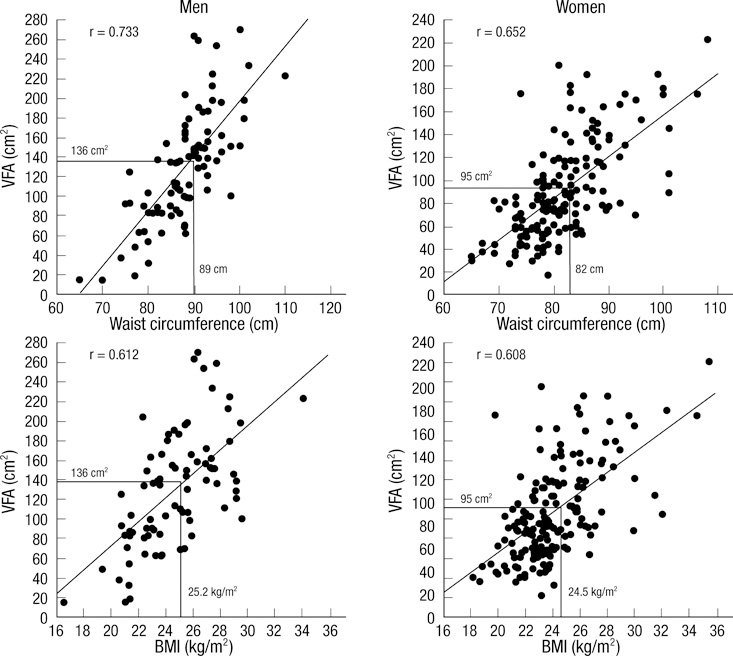
Fig. 1 Body mass index and waist circumference corresponding to the visceral fat area (VFA) cutoff values for men and women.
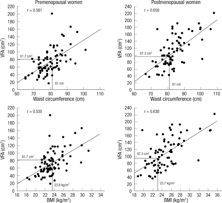
Fig. 2 Body mass index and waist circumference corresponding to the visceral fat area (VFA) cutoff values for pre- and post-menopausal women.
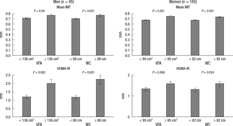
Fig. 3 Mean IMT and HOMA-IR according to the cutoff point of the visceral fat area (VFA) and waist circumference (WC) for men and women.
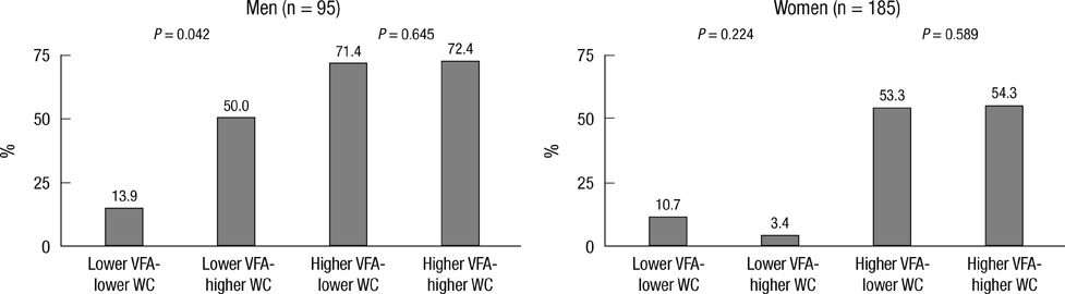
Fig. 4 Prevalence of metabolic syndrome according to the cutoff point of the visceral fat area (VFA) and waist circumference (WC) for men and women.
Cited by 2 articles
-
Relationship between Abdominal Fat Area Measured by Screening Abdominal Fat CT and Metabolic Syndrome in Asymptomatic Korean Individuals
Dae Woong Park, Noh Hyuck Park, Ji Yeon Park, Seon-Jeong Kim
J Korean Soc Radiol. 2017;77(1):1-8. doi: 10.3348/jksr.2017.77.1.1.Sex Differences of Visceral Fat Area and Visceral-to-Subcutaneous Fat Ratio for the Risk of Incident Type 2 Diabetes Mellitus
Eun Hee Kim, Hong-Kyu Kim, Min Jung Lee, Sung-Jin Bae, Jaewon Choe, Chang Hee Jung, Chul-Hee Kim, Joong-Yeol Park, Woo Je Lee
Diabetes Metab J. 2022;46(3):486-498. doi: 10.4093/dmj.2021.0095.
Reference
-
1. Gami AS, Witt BJ, Howard DE, Erwin PJ, Gami LA, Somers VK, Montori VM. Metabolic syndrome and risk of incident cardiovascular events and death: a systematic review and meta-analysis of longitudinal studies. J Am Coll Cardiol. 2007. 49:403–414.2. Després JP. Abdominal obesity as important component of insulin-resistance syndrome. Nutrition. 1993. 9:452–459.3. Carr DB, Utzschneider KM, Hull RL, Kodama K, Retzlaff BM, Brunzell JD, Shofer JB, Fish BE, Knopp RH, Kahn SE. Intra-abdominal fat is a major determinant of the National Cholesterol Education Program Adult Treatment Panel III criteria for the metabolic syndrome. Diabetes. 2004. 53:2087–2094.4. Pischon T, Boeing H, Hoffmann K, Bergmann M, Schulze MB, Overvad K, van der Schouw YT, Spencer E, Moons KG, Tjønneland A, Halkjaer J, Jensen MK, Stegger J, Clavel-Chapelon F, Boutron-Ruault MC, Chajes V, Linseisen J, Kaaks R, Trichopoulou A, Trichopoulos D, Bamia C, Sieri S, Palli D, Tumino R, Vineis P, Panico S, Peeters PH, May AM, Bueno-de-Mesquita HB, van Duijnhoven FJ, Hallmans G, Weinehall L, Manjer J, Hedblad B, Lund E, Agudo A, Arriola L, Barricarte A, Navarro C, Martinez C, Quirós JR, Key T, Bingham S, Khaw KT, Boffetta P, Jenab M, Ferrari P, Riboli E. General and abdominal adiposity and risk of death in Europe. N Engl J Med. 2008. 359:2105–2120.5. Alberti KG, Zimmet P, Shaw J. Metabolic syndrome: a new world-wide definition. A Consensus Statement from the International Diabetes Federation. Diabet Med. 2006. 23:469–480.6. Misra A, Vikram NK, Gupta R, Pandey RM, Wasir JS, Gupta VP. Waist circumference cutoff points and action levels for Asian Indians for identification of abdominal obesity. Int J Obes (Lond). 2006. 30:106–111.7. Huxley R, Barzi F, Lee CM, Lear S, Shaw J, Lam TH, Caterson I, Azizi F, Patel J, Suriyawongpaisal P, Oh SW, Kang JH, Gill T, Zimmet P, James PT, Woodward M. Waist circumference thresholds provide an accurate and widely applicable method for the discrimination of diabetes. Diabetes Care. 2007. 30:3116–3118.8. Janssen I, Katzmarzyk PT, Ross R. Waist circumference and not body mass index explains obesity-related health risk. Am J Clin Nutr. 2004. 79:379–384.9. Ross R, Léger L, Morris D, de Guise J, Guardo R. Quantification of adipose tissue by MRI: relationship with anthropometric variables. J Appl Physiol. 1992. 72:787–795.10. Thaete FL, Colberg SR, Burke T, Kelley DE. Reproducibility of computed tomography measurement of visceral adipose tissue area. Int J Obes Relat Metab Disord. 1995. 19:464–467.11. Kim SK, Kim HJ, Hur KY, Choi SH, Ahn CW, Lim SK, Kim KR, Lee HC, Huh KB, Cha BS. Visceral fat thickness measured by ultrasonography can estimate not only visceral obesity but also risks of cardiovascular and metabolic diseases. Am J Clin Nutr. 2004. 79:593–599.12. Pouliot MC, Després JP, Lemieux S, Moorjani S, Bouchard C, Tremblay A, Nadeau A, Lupien PJ. Waist circumference and abdominal sagittal diameter: best simple anthropometric indexes of abdominal visceral adipose tissue accumulation and related cardiovascular risk in men and women. Am J Cardiol. 1994. 73:460–468.13. Matthews DR, Hosker JP, Rudenski AS, Naylor BA, Treacher DF, Turner RC. Homeostasis model assessment: insulin resistance and β-cell function from fasting plasma glucose and insulin concentrations in man. Diabetologia. 1985. 28:412–419.14. Yanase T, Nasu S, Mukuta Y, Shimizu Y, Nishihara T, Okabe T, Nomura M, Inoguchi T, Nawata H. Evaluation of a new carotid intima-media thickness measurement by B-mode ultrasonography using an innovative measurement software, intimascope. Am J Hypertens. 2006. 19:1206–1212.15. Hanley JA, McNeil BJ. A method of comparing the areas under receiver operating characteristic curves derived from the same cases. Radiology. 1983. 148:839–843.16. Youden WJ. Index for rating diagnostic tests. Cancer. 1950. 3:32–35.17. Han JH, Park HS, Kim SM, Lee SY, Kim DJ, Choi WH. Visceral adipose tissue as a predictor for metabolic risk factors in the Korean population. Diabet Med. 2008. 25:106–110.18. Kim JA, Choi CJ, Yum KS. Cut-off values of visceral fat area and waist circumference: diagnostic criteria for abdominal obesity in a Korean population. J Korean Med Sci. 2006. 21:1048–1053.19. Oka R, Kobayashi J, Yagi K, Tanii H, Miyamoto S, Asano A, Hagishita T, Mori M, Moriuchi T, Kobayashi M, Katsuda S, Kawashiri MA, Nohara A, Takeda Y, Mabuchi H, Yamagishi M. Reassessment of the cutoff values of waist circumference and visceral fat area for identifying Japanese subjects at risk for the metabolic syndrome. Diabetes Res Clin Pract. 2008. 79:474–481.20. Brochu M, Starling RD, Tchernof A, Matthews DE, Garcia-Rubi E, Poehlman ET. Visceral adipose tissue is an independent correlate of glucose disposal in older obese postmenopausal women. J Clin Endocrinol Metab. 2000. 85:2378–2384.21. Bonora E, Micciolo R, Ghiatas AA, Lancaster JL, Alyassin A, Muggeo M, DeFronzo RA. Is it possible to derive a reliable estimate of human visceral and subcutaneous abdominal adipose tissue from simple anthropometric measurements? Metabolism. 1995. 44:1617–1625.22. Hyun YJ, Kim OY, Jang Y, Ha JW, Chae JS, Kim JY, Yeo HY, Paik JK, Lee JH. Evaluation of metabolic syndrome risk in Korean premenopausal women: not waist circumference but visceral fat. Circ J. 2008. 72:1308–1315.23. Seo JA, Kim BG, Cho H, Kim HS, Park J, Baik SH, Choi DS, Park MH, Jo SA, Koh YH, Han C, Kim NH. The cutoff values of visceral fat area and waist circumference for identifying subjects at risk for metabolic syndrome in elderly Korean: Ansan Geriatric (AGE) cohort study. BMC Public Health. 2009. 9:443.24. Enzi G, Gasparo M, Biondetti PR, Fiore D, Semisa M, Zurlo F. Subcutaneous and visceral fat distribution according to sex, age, and overweight, evaluated by computed tomography. Am J Clin Nutr. 1986. 44:739–746.25. Jeppesen J, Hansen TW, Rasmussen S, Ibsen H, Torp-Pederson C, Madsbad S. Insulin resistance, the metabolic syndrome, and risk of incident cardiovascular disease: a population-based study. J Am Coll Cardiol. 2007. 49:2112–2119.26. Bots ML, Hoes AW, Koudstaal PJ, Hofman A, Grobbee DE. Common carotid intima-media thickness and risk of stroke and myocardial infarction: the Rotterdam Study. Circulation. 1997. 96:1432–1437.27. Tabata S, Yoshimitsu S, Hamachi T, Abe H, Ohnaka K, Kono S. Waist circumference and insulin resistance: a cross-sectional study of Japanese men. BMC Endocr Disord. 2009. 9:1.28. Park K, Lee DH, Erickson DJ, Himes JH, Shikany JM, Jacobs DR Jr. Association of long-term change in waist circumference with insulin resistance. Obesity. 2010. 18:370–376.29. Hassinen M, Lakka TA, Komulainen P, Haapala I, Nissinen A, Rauramaa R. Association of waist and hip circumference with 12-year progression of carotid intima-media thickness in elderly women. Int J Obes (Lond). 2007. 31:1406–1411.
- Full Text Links
-
- Actions
-
Cited
- CITED
-
- Close
- Share
-
- Similar articles
-
- The Association of Visceral Fat Area with Anthropometric Variables and its Risk for Metabolic Syndrome
- Visceral Obesity
- Relationship of Metabolic Syndrome and Truncal Obesity Measured by Dual Energy X-ray Absorptiometry and Computed Tomography in Obese Women
- The Accuracy of the Assessment of Visceral Obesity by InBody 4.0 and Waist Circumference
- Cut-off Values of Visceral Fat Area and Waist Circumference: Diagnostic Criteria for Abdominal Obesity in a Korean Population
