Myoepitheliomas of the Soft Palate: Helical CT Findings in Two Patients
- Affiliations
-
- 1Department of Radiology, Eulji Medical Center, Eulji University School of Medicine, Seoul, Korea. khs46359@eulji.ac.kr
- 2Department of Pathology, Eulji University School of Medicine, Daejeon, Korea.
- 3Department of Otorhinolaryngology, Eulji Medical Center, Eulji University School of Medicine, Daejeon, Korea.
- KMID: 1089445
- DOI: http://doi.org/10.3348/kjr.2007.8.6.552
Abstract
- We describe the enhancement patterns of myoepithelioma in two patients with a soft palate mass. In the first case, helical CT revealed a faintly enhancing mass. Histologically, the tumor was composed of plasmacytoid cells in a background of rich myxoid stroma. Immunostaining for CD34 showed scanty blood vessels. In the second case, helical CT revealed an intensely enhancing mass. Histologically, the mass was a cellular tumor with fibrous stroma. Immunostaining for CD34 also showed frequent blood vessels.
MeSH Terms
-
Aged
Contrast Media/administration & dosage
Diagnosis, Differential
Female
Humans
Iohexol/analogs & derivatives/diagnostic use
Male
Middle Aged
Myoepithelioma/*diagnosis/surgery
Palatal Neoplasms/*diagnosis/surgery
Palate, Soft/*radiography/surgery
Radiographic Image Enhancement/methods
Rare Diseases
Tomography, Spiral Computed/*methods
Figure
-
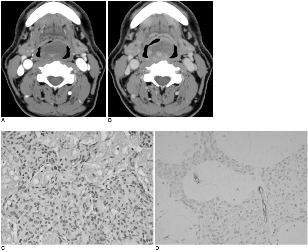
Fig. 1 Case 1. A. An early phase axial CT scan shows a well-demarcated soft palate tumor showing faint enhancement. B. A delayed axial CT scan shows heterogenous, nodular enhancement of the tumor. C. A microscopic view (Hematoxylin & Eosin staining ×400) of the myoepithelioma shows plasmacytoid cells in the background of myxoid stroma. D. Immunostaining for CD34 (×400) shows scanty blood vessels.

Fig. 2 Case 2. A. An early phase coronal CT scan shows an intensely enhancing soft palate tumor. B. A delayed axial CT scan shows persistent homogenous enhancement of the tumor. C. A microscopic view (Hematoxylin & Eosin staining ×400) of the myoepithelioma shows a cellular tumor composed of spindle cells. D. Immunostaining for CD34 (×400) shows frequent blood vessels.
Reference
-
1. Onbas O, Karasen RM, Gursan N, Kantarci M, Alper F, Okur A. Giant myoepithelioma of the face: MDCT with 2D and 3D images. AJR Am J Roentgenol. 2006. 187:W418–W419.2. Hiwatashi A, Matsumoto S, Kamoi I, Yamashita H, Nakashima A. Imaging features of myoepithelioma arising from the hard palate. A case report. Acta Radiol. 2000. 41:417–419.3. Kanazawa H, Furuya T, Watanabe T, Kato J. Plasmacytoid myoepithelioma of the palate. J Oral Maxillofac Surg. 1999. 57:857–860.4. Turgut S, Cekic A, Ergül G, Aksoy F, Seckin S, Ozdem C. Myoepithelioma of the parotid gland: A report of two cases. Ear Nose Throat J. 2001. 80:155–158.5. Spreer J, Krahe T, Jung G, Lackner K. Spiral versus conventional CT in routine examinations of the neck. J Comput Assist Tomogr. 1995. 19:905–910.6. Choi DS, Na DG, Byun HS, Ko YH, Kim CK, Cho JM. Salivary gland tumors: evaluation with two-phase helical CT. Radiology. 2000. 214:231–236.7. Lee DK, Chung KW, Baek CH, Jeong HS, Ko YH, Son YI. Basal cell adenoma of the parotid gland. characteristics of 2-phase helical computed tomography and magnetic resonance imaging. J Comput Assist Tomogr. 2005. 29:884–888.8. Mukherji SK. Som PM, Curtin HD, editors. Pharynx. Head and neck imaging. 2003. 4th ed. St. Louis: Mosby;1465–1520.
- Full Text Links
-
- Actions
-
Cited
- CITED
-
- Close
- Share
-
- Similar articles
-
- Histopathologic study of soft palate muscles in cleft palate (II)>
- Prosthetic rehabilitation of soft palate resection edentulous patient with maxillary obturator
- Central Lucency of Pelvic Phleboliths: Comparison of Plain Radiographs and Noncontrast Helical CT
- CT findings of polymorphic reticulosis: 5 case reports
- Diagnostic Accuracy and Usefulness of Three Dimensional Image of Helical CT in Maxillofacial Fractures
